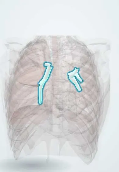
Lobar bronchi !
The lobar bronchi are the airways that branch from each main bronchus to carry air to a single lobe: three on the right, two on the left.
Official name: Lobar bronchus
Opens in the Durgical app, no sign-up needed.
What it is
The branches of the main bronchi to each lobe: three on the right (with the intermediate bronchus) and two on the left.
What it does
Carry air to each lobe.
Key facts
- Where
- Right: upper (eparterial, above pulmonary artery), middle, lower; left: upper and lower
- Blood supply
- Bronchial arteries (left from thoracic aorta; right usually from 3rd right posterior intercostal artery)
- Nerve
- Pulmonary plexus: vagus (bronchoconstriction, secretion); upper thoracic sympathetic (bronchodilation)
- Function
- Conduct air to each lobe (secondary bronchi): 3 on right, 2 on left
Relations
- Course
- Right: the upper lobe bronchus leaves first; the bronchus intermedius continues down and gives the middle lobe bronchus forward and the lower lobe bronchus; left: after passing under the aortic arch the main bronchus divides into upper and lower lobe bronchi
- In front
- Right: the right pulmonary artery crosses in front of the bronchus intermedius; at both hila the superior pulmonary veins lie in front of the bronchi
- Above
- Left: the left pulmonary artery arches over the upper lobe bronchus and then descends behind and lateral to the lower lobe bronchus
How to find it
Found at bronchoscopy. On the right, the upper lobe opening is on the lateral wall just beyond the carina; lower down, the middle lobe opening faces forward and the superior segment of the lower lobe opens backward at about the same level. On the left, a secondary carina separates the upper and lower lobe bronchi.
On imaging
Lobar collapse follows the blocked bronchus. Right upper lobe: the horizontal fissure lifts; with a central mass it forms a reversed S (Golden S sign). Left upper lobe: a veil-like haze over the left chest, sometimes with a crescent of air beside the aortic knob (Luftsichel sign). Lower lobes: a triangle beside the spine with the hilum pulled down.
Common problems
- Narrowing after a long time with a breathing tube.
- Inhaled objects, especially in children.
How it is diagnosed
CT, bronchoscopy.
Treatment
- Bronchoscopy to remove objects; stents or surgery for narrowings.
Clinical notes
- A central tumour blocking a lobar bronchus causes collapse and post-obstructive pneumonia; pneumonia that keeps returning in the same lobe raises this concern
- In a lobectomy the lobar bronchus, artery and vein are divided separately at the hilum and the bronchial stump is closed; a stump leak (bronchopleural fistula) is a feared complication
- Endobronchial tuberculosis can scar and narrow a lobar bronchus, leaving collapse or bronchiectasis beyond it
Anatomical variants
- Tracheal bronchus: the right upper lobe bronchus, or one of its segmental branches, arises from the trachea above the carina; usually an incidental finding, sometimes linked to recurrent right upper lobe infection
Development
Each main bronchial bud divides into secondary (lobar) buds, three on the right and two on the left; each one becomes a lobar bronchus and the lobe it ventilates.
Related conditions
- Cystic fibrosis
- Atelectasis
- Acute bronchitis
- Allergic bronchopulmonary aspergillosis (ABPA)
- Asthma
- Lung cancer
- Bronchiectasis
Test yourself
Think of the answer, then tap the question to see it.
Why is the right middle lobe bronchus prone to repeated or chronic collapse (middle lobe syndrome)?
It is long and narrow and ringed by lymph nodes, so it is easily compressed or blocked.
Which right lobar bronchus is called eparterial?
The right upper lobe bronchus, which arises above the pulmonary artery, almost at once from the right main bronchus.
What does pneumonia that keeps returning in the same lobe raise concern for?
A central tumour blocking the lobar bronchus, causing collapse and post-obstructive pneumonia beyond it.
What is the Golden S sign on a chest X-ray?
A reversed S outline of the lifted horizontal fissure when collapse of the right upper lobe is caused by a central mass.
Educational information, not medical advice. For any symptom, see a health professional.
Its names
- Official Latin (TA2)
- bronchus lobaris TA2 3229
- Official Spanish (DeCS)
- Bronquios lobares DeCS D001980
- English (TA2)
- Lobar bronchus
- Also called
- secondary bronchus
Related
Sources: Terminologia Anatomica (TA2); DeCS; Anatomy, Thorax, Tracheobronchial Tree; Anatomy, Thorax, Bronchial; 22.7 Embryonic Development of the Respiratory System (OpenStax (Rice University), 2022) · adapted, CC BY 4.0; Anatomy of the Human Body (20th ed.): The Respiratory Apparatus (Lea & Febiger (Gray's Anatomy, 1918), 1918) · adapted, Public domain.
Spotted a mistake?We are always improving for the community. Tell us what is wrong and our team will review it.Report a mistake
Representative model, for educational purposes only.