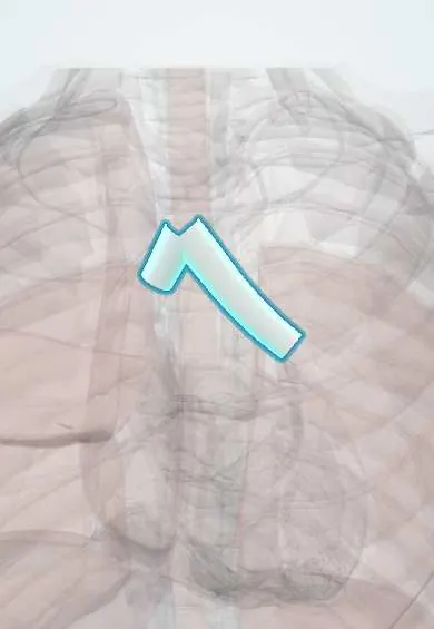
Main bronchi !
The main bronchi are the two airways that branch from the windpipe, one into each lung; the right is wider and steeper than the left.
Official name: Right main bronchus
Opens in the Durgical app, no sign-up needed.
What it is
The two first branches of the trachea, one to each lung. The right is shorter, wider and more vertical.
What it does
Carry air into each lung. Inhaled objects usually go to the right side.
Key facts
- Where
- From carina (about T4–T5); right shorter, wider, more vertical; left passes under aortic arch, in front of esophagus
- Blood supply
- Bronchial arteries (left from thoracic aorta; right usually from 3rd right posterior intercostal artery)
- Nerve
- Pulmonary plexus: vagus (bronchoconstriction, secretion); upper thoracic sympathetic (bronchodilation)
- Function
- Conduct air to each lung; inhaled foreign bodies enter the right one more often
Relations
- Above
- Right: the arch of the azygos vein, on its way to the superior vena cava; left: the aortic arch and, closer to the bronchus, the left pulmonary artery as it crosses over it
- In front
- Right: the superior vena cava and the right pulmonary artery; left: the left pulmonary artery and, lower at the hilum, the pulmonary veins
- Behind
- Right: the azygos vein before it arches forward; left: the esophagus and the descending thoracic aorta
- Below
- The subcarinal lymph nodes in the angle between the two bronchi, above the left atrium
How to find it
Not palpable. The carina projects at the sternal angle. At bronchoscopy the right main bronchus continues almost straight down from the trachea, while the left leaves at a wider angle and runs more horizontally under the aortic arch. On CT both are followed from the carina to the hila.
On imaging
On a frontal chest X-ray both air columns can be traced through the heart shadow; the left runs more horizontally under the aortic knob. A widened angle between them points to an enlarged left atrium or subcarinal nodes. Coronal CT reformats show each bronchus in its full length.
Common problems
- Narrowing after a long time with a breathing tube.
- Inhaled objects, especially in children.
How it is diagnosed
CT, bronchoscopy.
Treatment
- Bronchoscopy to remove objects; stents or surgery for narrowings.
Clinical notes
- Tube placement is checked by listening over both sides of the chest and on a chest X-ray; unequal breath sounds suggest a tube in one main bronchus
- Double-lumen tubes for one-lung ventilation are usually placed in the left main bronchus, because on the right the upper lobe bronchus arises so close to the carina that it is easily blocked
- Blunt chest trauma can tear a main bronchus close to the carina, giving a persistent air leak, pneumomediastinum and a lung that will not re-expand
Development
The two main bronchi come from the paired bronchial (lung) buds at the lower end of the laryngotracheal tube in week 4 to 5. From the start the right bud is the larger and more in line with the trachea, the same asymmetry seen in the adult.
Memory aid
Inhale a bite, it goes down the right: the right main bronchus is wider and more in line with the trachea.
Related conditions
- Anaphylaxis
- Organophosphate and carbamate poisoning
- Foreign-body airway obstruction (choking)
- Pneumocystis pneumonia (PCP)
- Influenza (flu)
- Pertussis (whooping cough)
- Foreign body aspiration in children
- Meconium aspiration syndrome
- Aspiration pneumonia and pneumonitis
- Lung abscess
- Acute bronchitis
- Asthma
- Lung cancer
Test yourself
Think of the answer, then tap the question to see it.
Which main bronchus does an endotracheal tube pushed too far usually enter, and what is the result?
The right main bronchus; the left lung is not ventilated, left breath sounds are lost and the left lung can collapse.
How do the right and left main bronchi differ in shape?
The right is shorter (about 2.5 cm), wider and more vertical; the left is about twice as long and runs more horizontally under the aortic arch.
Which vessels hook over the right and the left main bronchus?
The arch of the azygos vein over the right main bronchus; the aortic arch over the left.
Why are double-lumen tubes for one-lung ventilation usually placed in the left main bronchus?
On the right the upper lobe bronchus arises so close to the carina that the tube easily blocks it.
Educational information, not medical advice. For any symptom, see a health professional.
Its names
- Official Latin (TA2)
- bronchus principalis dexter; bronchus principalis sinister TA2 3237
- Official Spanish (DeCS)
- Bronquios principales derecho e izquierdo DeCS D001980
- English (TA2)
- Right main bronchus
Related
Sources: Terminologia Anatomica (TA2); DeCS; Anatomy, Thorax, Tracheobronchial Tree; Anatomy, Thorax, Bronchial; 22.7 Embryonic Development of the Respiratory System (OpenStax (Rice University), 2022) · adapted, CC BY 4.0; Anatomy of the Human Body (20th ed.): The Respiratory Apparatus (Lea & Febiger (Gray's Anatomy, 1918), 1918) · adapted, Public domain.
Spotted a mistake?We are always improving for the community. Tell us what is wrong and our team will review it.Report a mistake
Representative model, for educational purposes only.