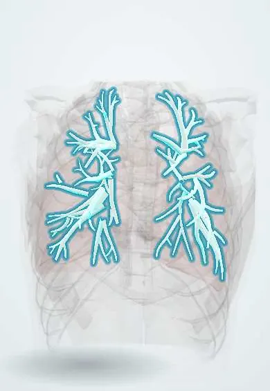
Segmental bronchi !
The segmental bronchi are the airways that carry air to each lung segment; doctors number them to find their way during bronchoscopy.
Official name: Segmental bronchus
Opens in the Durgical app, no sign-up needed.
What it is
The branches to each bronchopulmonary segment, numbered B1 to B10.
What it does
Carry air to each segment; bronchoscopists navigate by these numbers.
Key facts
- Where
- Tertiary bronchi numbered B1–B10: 10 on right, 8–10 on left
- Blood supply
- Bronchial arteries (left from thoracic aorta; right usually from 3rd right posterior intercostal artery)
- Nerve
- Pulmonary plexus: vagus (bronchoconstriction, secretion); upper thoracic sympathetic (bronchodilation)
- Function
- Each aerates one bronchopulmonary segment; landmarks for bronchoscopy and segmentectomy
Relations
- Course
- Each enters the centre of its bronchopulmonary segment with the segmental branch of the pulmonary artery alongside, then divides into subsegmental bronchi and bronchioles; the cartilage is in irregular plates, not rings
How to find it
Found at bronchoscopy, where each opening is named by its number. Postural drainage places the target segmental bronchus so that gravity carries secretions toward the trachea, which is why the segment map matters in chest physiotherapy.
On imaging
On thin-section CT each segmental bronchus runs beside its artery, both of about the same calibre; a bronchus clearly wider than its artery (signet ring sign) means bronchiectasis. Segmental consolidation or infarction is wedge-shaped, with its apex toward the hilum.
Common problems
- Narrowing after a long time with a breathing tube.
- Inhaled objects, especially in children.
How it is diagnosed
CT, bronchoscopy.
Treatment
- Bronchoscopy to remove objects; stents or surgery for narrowings.
Clinical notes
- Segmentectomy removes one bronchopulmonary segment with its own bronchus and artery, sparing lung in small peripheral tumours or when lung reserve is low
- Lung abscess after aspiration typically sits in the posterior segment of the right upper lobe or the superior segment of the right lower lobe
- Post-tuberculous bronchiectasis favours the apical and posterior segments of the upper lobes, a pattern seen often where tuberculosis is still common, as in Colombia
Development
By about week 7 the lobar buds have branched into tertiary (segmental) buds, ten on the right and eight to ten on the left; each one, with the mesenchyme around it, becomes a bronchopulmonary segment. Branching then continues well beyond this level until the respiratory bronchioles form.
Memory aid
Right lung in order, B1 to B10: A P A, L M, S M A L P (Apical, Posterior, Anterior; Lateral, Medial; Superior, Medial, Anterior, Lateral, Posterior basal).
Related conditions
- Hydrocarbon ingestion and aspiration
- Aspergillosis
- Bronchiolitis (RSV)
- Meconium aspiration syndrome
- Bronchopulmonary dysplasia
- Cystic fibrosis
- Hypersensitivity pneumonitis
- Allergic bronchopulmonary aspergillosis (ABPA)
- Asthma
- Chronic obstructive pulmonary disease (COPD)
- Bronchiectasis
Test yourself
Think of the answer, then tap the question to see it.
How are the segmental bronchi of the right lung numbered from B1 to B10?
Upper lobe apical, posterior, anterior; middle lobe lateral, medial; lower lobe superior, then medial, anterior, lateral and posterior basal.
What is the signet ring sign on thin-section CT of a segmental bronchus?
A bronchus clearly wider than its accompanying artery; it means bronchiectasis.
Which segments do post-tuberculous bronchiectasis favour?
The apical and posterior segments of the upper lobes.
Educational information, not medical advice. For any symptom, see a health professional.
Its names
- Official Latin (TA2)
- bronchus segmentalis TA2 3230
- Official Spanish (DeCS)
- Bronquios segmentarios DeCS D001980
- English (TA2)
- Segmental bronchus
- Also called
- tertiary bronchus
Related
Sources: Terminologia Anatomica (TA2); DeCS; Anatomy, Thorax, Tracheobronchial Tree; Anatomy, Thorax, Bronchial; 22.7 Embryonic Development of the Respiratory System (OpenStax (Rice University), 2022) · adapted, CC BY 4.0.
Spotted a mistake?We are always improving for the community. Tell us what is wrong and our team will review it.Report a mistake
Representative model, for educational purposes only.