Conditions · Ophthalmology and ENT
Central retinal vein occlusion !
A clot blocks the main vein draining the retina, causing sudden painless blurred vision in one eye. It is linked to high blood pressure and glaucoma.
Updated 2026-11-01
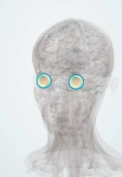
Structures involved
Tap one to see what it is and open it in 3D.
Why it happens
- Thrombosis and Virchow's triad
- Hypercoagulable states: inherited and acquired
- Retina: the ten layers, rods and cones
What it is
Blockage of the central retinal vein, usually at or behind the lamina cribrosa of the optic disc, which causes venous congestion, haemorrhage and swelling throughout the retina and painless loss of vision in one eye.
Who gets it
Retinal vein occlusion as a group (branch and central) is the second most common retinal vascular disease after diabetic retinopathy; the central form mainly affects people over 55. In Latin America it is seen mostly in older adults with hypertension, diabetes or glaucoma.
Causes
- Thrombosis of the central retinal vein, where it shares a sheath with a stiff, atherosclerotic central retinal artery
- Hyperviscosity or hypercoagulable states in younger patients
- Inflammatory vessel disease (vasculitis) in some cases
Risk factors
- Age over 55
- Arterial hypertension (the most common association)
- Diabetes and dyslipidaemia
- Open-angle glaucoma or raised intraocular pressure
- Thrombophilia, antiphospholipid syndrome, polycythaemia, myeloma; oral contraceptives in young women
Pathophysiology
The obstructed vein raises pressure in the retinal capillaries, so blood and fluid leak into the retina and macula. Poor outflow reduces arterial inflow, causing retinal ischaemia. The ischaemic retina releases VEGF, which drives macular oedema and abnormal new vessels on the iris and angle, which can close the angle and cause neovascular glaucoma.
Classification
- Non-ischaemic (perfused): better vision, mild or no relative afferent pupillary defect; most common
- Ischaemic (non-perfused): severe vision loss, marked afferent pupillary defect, wide capillary non-perfusion; high risk of neovascularisation
- Related: branch retinal vein occlusion and hemiretinal vein occlusion
Symptoms and signs
- Sudden or rapidly developing painless blurred vision in one eye
- Sometimes milder, fluctuating blurring on waking
- Pain and redness appear only later if neovascular glaucoma develops
Diagnosis
- Clinical
- Visual acuity, pupils (relative afferent pupillary defect suggests ischaemia), intraocular pressure and gonioscopy, and dilated fundoscopy: dilated tortuous veins and haemorrhages in all four quadrants, the 'blood and thunder' or 'stormy sunset' fundus, with disc and macular swelling and cotton-wool spots.
- Tests
- Blood pressure, glucose or HbA1c, lipids, complete blood count, ESR, renal function. In young or bilateral cases: thrombophilia screen, antiphospholipid antibodies, homocysteine and protein electrophoresis.
- Imaging
- Optical coherence tomography measures macular oedema; fluorescein angiography shows the extent of capillary non-perfusion (ischaemic vs non-ischaemic).
Differential diagnosis
- Diabetic retinopathy
- Hypertensive retinopathy
- Ocular ischaemic syndrome (carotid stenosis)
- Central retinal artery occlusion
- Papilloedema
Goal of treatment
Treat macular oedema to recover vision, prevent and treat neovascular glaucoma, and control the systemic risk factors that caused the occlusion.
Treatment
- Procedure
- Intravitreal anti-VEGF injections are first-line for macular oedema; intravitreal corticosteroids are an alternative.
- Procedure
- Panretinal laser photocoagulation when new vessels appear on the iris, angle or retina.
- Medical
- Control of hypertension, diabetes, lipids and intraocular pressure; treatment of any blood or clotting disorder found.
- Surgical
- Vitrectomy for non-clearing vitreous haemorrhage; glaucoma surgery for refractory neovascular glaucoma.
- Follow-up
- Frequent visits (often monthly in ischaemic cases for the first months) with gonioscopy to detect iris neovascularisation early.
Complications
- Chronic macular oedema and permanent vision loss
- Iris neovascularisation and neovascular glaucoma (classically within about 3 months)
- Vitreous haemorrhage
- Occlusion in the other eye
Prognosis
Final vision depends largely on vision at presentation and on whether the occlusion is ischaemic. Non-ischaemic cases often improve, though some convert to ischaemic; ischaemic cases have poor vision and need close watch for neovascular glaucoma.
Prevention
- Control of blood pressure, diabetes and lipids
- Regular screening and treatment of glaucoma
Test yourself
Think of the answer, then tap the question to see it.
What is the classic fundus appearance of central retinal vein occlusion?
Dilated, tortuous veins with haemorrhages in all four quadrants, disc swelling and cotton-wool spots ('blood and thunder' fundus).
Which systemic disease is most strongly associated with central retinal vein occlusion?
Arterial hypertension; diabetes, dyslipidaemia and glaucoma are other key associations.
What feared complication follows ischaemic central retinal vein occlusion within about 3 months?
Iris neovascularisation leading to neovascular glaucoma (the '90-day glaucoma').
What is first-line treatment for macular oedema due to retinal vein occlusion?
Intravitreal anti-VEGF injections.
Related conditions
- Arterial hypertension
- Open-angle glaucoma
- Type 2 diabetes mellitus
- Central retinal artery occlusion
- Hypertensive retinopathy
- Diabetic retinopathy
- Polycythaemia vera
- Antiphospholipid syndrome
Educational information, not medical advice. For any symptom, see a health professional.
Sources: Central Retinal Vein Occlusion (EyeWiki (American Academy of Ophthalmology), 2025).
Spotted a mistake?We are always improving for the community. Tell us what is wrong and our team will review it.Report a mistake
Representative model, for educational purposes only.