Conditions · Systemic conditions
Leptospirosis !
Leptospirosis is a bacterial infection caught from water or soil soiled by the urine of infected animals, mainly rats; it ranges from flu-like to severe.
Updated 2026-10-29
Structures involved
Tap one to see what it is and open it in 3D.
Microorganisms that cause it
In the clinical exam
Why it happens
What it is
A zoonotic bacterial infection caused by Leptospira species, acquired through contact with water or soil contaminated by the urine of infected animals, especially rodents. It ranges from a flu-like illness to severe Weil disease.
Who gets it
Common in tropical and subtropical regions, with outbreaks after heavy rain and floods. In Colombia and much of Latin America it is a recognised public-health problem and a notifiable disease, affecting urban slums as well as farm and livestock workers.
Causes
- Pathogenic Leptospira species (spirochaetes), most often L. interrogans
- Urine of infected rodents (main reservoir), dogs, cattle and pigs
- Entry through cuts, abraded skin or mucous membranes (eyes, mouth, nose)
- Exposure to floodwater, mud, sewers and contaminated rivers or ponds
Risk factors
- Floods and heavy rains
- Living in areas with poor sanitation and many rodents
- Farming, rice growing, livestock handling and slaughterhouse work
- Sewer and garbage workers, veterinarians
- Freshwater recreation, adventure sports and military field work
- Walking barefoot or with skin wounds in contaminated water
Pathophysiology
Leptospires enter through skin or mucosa, spread in the blood and reach almost every organ (leptospiraemic phase). They damage small-vessel endothelium, causing vasculitis, capillary leak and bleeding. An immune phase follows as antibodies appear; in severe cases liver dysfunction, acute kidney injury (often non-oliguric, with potassium loss) and pulmonary haemorrhage develop.
Classification
- Anicteric leptospirosis: mild, self-limited, flu-like illness (most cases)
- Icteric leptospirosis (Weil disease): jaundice, acute kidney injury and bleeding
- Severe pulmonary haemorrhage syndrome
- Classically biphasic: septicaemic phase then immune phase, although phases often overlap
Symptoms and signs
- Sudden high fever, chills and severe headache
- Intense muscle pain, typically in the calves and lower back
- Conjunctival suffusion (red eyes without discharge)
- Nausea, vomiting, abdominal pain, diarrhoea
- Jaundice in severe forms
- Reduced urine output, cough, bloody sputum or rash in some patients
Diagnosis
- Clinical
- Suspected in fever with calf pain and red eyes after exposure to floods, rodents or animals. A history of exposure in the weeks before is key.
- Tests
- Blood or urine PCR early in illness; IgM ELISA and the microscopic agglutination test (MAT, the reference serologic test) from the end of the first week. Labs may show thrombocytopenia, raised creatine kinase, high bilirubin with only modestly raised transaminases, raised creatinine and low potassium.
- Imaging
- Chest X-ray or CT for patchy or diffuse infiltrates when pulmonary haemorrhage is suspected; ultrasound if biliary or renal problems need to be assessed.
Differential diagnosis
- Dengue
- Malaria
- Viral hepatitis
- Typhoid fever
- Bacterial sepsis
- Rickettsial infection
Goal of treatment
Clear the infection early with antibiotics and support failing organs (kidney, lung, circulation) to prevent death in severe forms.
Treatment
- Medical
- Mild disease: oral antibiotics, usually tetracyclines (doxycycline) or aminopenicillins, started on clinical suspicion without waiting for confirmation.
- Medical
- Severe disease: intravenous penicillin or third-generation cephalosporins in hospital.
- Acute
- Supportive care: fluids, electrolyte replacement (especially potassium), oxygen and transfusion of platelets or blood products when needed.
- Procedure
- Renal replacement therapy (dialysis) for severe acute kidney injury and mechanical ventilation for pulmonary haemorrhage.
- Follow-up
- Close monitoring of urine output, creatinine, potassium, bilirubin, platelets and oxygenation; a Jarisch–Herxheimer reaction may follow the first antibiotic dose.
Drugs for this condition
Complications
- Acute kidney injury
- Severe pulmonary haemorrhage and respiratory failure
- Liver dysfunction and jaundice
- Myocarditis and arrhythmias
- Bleeding and shock
- Uveitis weeks to months later
Prognosis
Most infections are mild and recover fully. Weil disease and pulmonary haemorrhage carry a high mortality, which falls with early antibiotics, dialysis and intensive care. Kidney function usually recovers in survivors.
Prevention
- Rodent control and better sanitation, drainage and waste collection
- Protective boots and gloves for exposed workers and during flood clean-up
- Avoiding swimming or wading in possibly contaminated water, especially with skin wounds
- Vaccination of livestock and dogs where available
- Antibiotic prophylaxis for selected high-risk exposures, according to local guidelines
Test yourself
Think of the answer, then tap the question to see it.
What is the main reservoir of leptospirosis and how is it transmitted to humans?
Rodents; humans are infected through skin cuts or mucosa exposed to water or soil contaminated with infected animal urine.
What is Weil disease?
The severe icteric form of leptospirosis with jaundice, acute kidney injury and bleeding.
Which two physical findings strongly suggest leptospirosis in a febrile patient after a flood?
Conjunctival suffusion (red eyes without discharge) and intense calf muscle pain.
What is the reference serologic test for leptospirosis?
The microscopic agglutination test (MAT).
What kind of kidney injury is typical in leptospirosis?
Often non-oliguric acute kidney injury with potassium loss (hypokalaemia).
Related conditions
Educational information, not medical advice. For any symptom, see a health professional.
Sources: Leptospirosis (StatPearls (NCBI Bookshelf), 2024).
Spotted a mistake?We are always improving for the community. Tell us what is wrong and our team will review it.Report a mistake
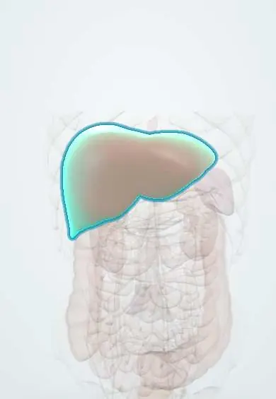
Representative model, for educational purposes only.