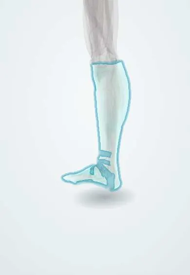
Cellulitis of the leg !
Cellulitis of the leg is a bacterial infection of the deep skin and fat beneath; it causes spreading redness, warmth, swelling and pain.
Updated 2026-10-29
Structures involved
Tap one to see what it is and open it in 3D.
Microorganisms that cause it
Why it happens
- Patterns of acute inflammation: serous, fibrinous, purulent, abscess, ulcer
- Vascular changes in acute inflammation: vasodilation, leakage, exudate vs transudate
- Inflammation: the cardinal signs and its five steps
- Skin: epidermis layers, dermis, hypodermis
What it is
Acute bacterial infection of the deep dermis and subcutaneous tissue of the leg, with spreading redness, warmth, swelling and pain.
Who gets it
A common reason for emergency visits and hospital admission, especially in adults with obesity, leg swelling or diabetes. In warm climates insect bites and fungal infection between the toes are frequent entry points.
Causes
- Beta-haemolytic streptococci (most non-purulent cases)
- Staphylococcus aureus, including MRSA, especially with pus or abscess
- Other organisms after water exposure, bites or in immunocompromised patients
Risk factors
- Breaks in the skin: wounds, ulcers, insect bites
- Tinea pedis (athlete’s foot)
- Lymphoedema or chronic venous insufficiency
- Obesity
- Diabetes
- Previous cellulitis
Pathophysiology
Bacteria enter through a break in the skin barrier and spread through the dermis and subcutaneous fat. Bacterial toxins and the immune response cause the redness, heat, oedema and pain. Erysipelas is a more superficial form involving the upper dermis and lymphatics.
Classification
- Non-purulent (usually streptococcal) versus purulent (often staphylococcal)
- Severity: mild, moderate or severe with systemic signs
- Erysipelas: superficial, sharply raised border
Symptoms and signs
- Red, warm, swollen and tender skin, usually on one leg
- Poorly defined edge (erysipelas has a raised, sharp edge)
- Fever and malaise
- Tender, enlarged inguinal lymph nodes
- Blisters in some cases
Diagnosis
- Clinical
- Clinical diagnosis from the appearance; outlining the border helps follow spread. Look for the entry point, such as tinea between the toes.
- Tests
- Blood count and inflammatory markers in moderate or severe cases; blood cultures if systemically unwell; culture of pus when present.
- Imaging
- Ultrasound to look for an abscess; Doppler if deep vein thrombosis is possible; CT or MRI only if a deep infection is suspected, without delaying surgery.
Differential diagnosis
- Deep vein thrombosis
- Stasis dermatitis
- Necrotising fasciitis
- Gout
- Contact dermatitis
- Erythema nodosum
Goal of treatment
Cure the infection, treat the entry point and prevent spread, complications and recurrence.
Treatment
- Medical
- Oral antibiotics active against streptococci for mild cases, adding staphylococcal or MRSA cover for purulent infection; intravenous antibiotics when severe.
- Conservative
- Elevation of the leg and treatment of oedema, tinea pedis or ulcers.
- Procedure
- Incision and drainage of any abscess.
- Acute
- Urgent surgical debridement and broad-spectrum antibiotics if necrotising fasciitis is suspected.
Drugs for this condition
- Amoxicillin + clavulanic acid
- Ampicillin + sulbactam
- Cefalexin
- Cefazolin
- Cefepime
- Ceftaroline
- Cloxacillin
- Dicloxacillin
- Oxacillin
- Nafcillin
- Flucloxacillin
- Daptomycin
- Dalbavancin
- Tedizolid
- Clindamycin
Complications
- Abscess
- Necrotising fasciitis
- Sepsis
- Recurrent cellulitis
- Lymphoedema from lymphatic damage
Prognosis
Most cases resolve with antibiotics within days. Recurrence is common when oedema, tinea or skin breaks persist.
Prevention
- Treating athlete’s foot and dry, cracked skin
- Controlling leg oedema with compression
- Early care of wounds and insect bites
Test yourself
Think of the answer, then tap the question to see it.
How does erysipelas differ from cellulitis?
Erysipelas is more superficial (upper dermis and lymphatics) with a raised, sharply defined border; cellulitis is deeper with a poorly defined edge.
Which bacteria usually cause non-purulent cellulitis?
Beta-haemolytic streptococci, mainly group A; Staphylococcus aureus is typical when there is pus.
Which finding should raise suspicion of necrotising fasciitis?
Pain out of proportion to the skin findings, rapid spread, bullae, crepitus or signs of sepsis.
Which common skin condition between the toes is a frequent entry point for leg cellulitis?
Tinea pedis (athlete’s foot).
Related conditions
- Diabetic foot
- Varicose veins
- Osteomyelitis
- Deep vein thrombosis
- Acute compartment syndrome
- Gout
- Chronic venous insufficiency
- Venous leg ulcer
Educational information, not medical advice. For any symptom, see a health professional.
Sources: Cellulitis (StatPearls (NCBI Bookshelf), 2025).
Spotted a mistake?We are always improving for the community. Tell us what is wrong and our team will review it.Report a mistake
Representative model, for educational purposes only.