Lymphoedema !
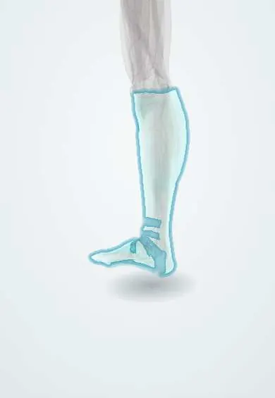
Lymphoedema is long-lasting swelling of a limb because the lymphatic system cannot drain fluid from the tissues.
Updated 2026-10-29
Structures involved
Tap one to see what it is and open it in 3D.
- Crural fascia
- Fascia lata
- Great saphenous vein
- Top-of-foot veins
- Second toe
- Inguinal ligament
- Femoral vein
Microorganisms that cause it
In immunology
In histology
In the clinical exam
In physiology
Why it happens
What it is
Chronic swelling of a limb caused by build-up of protein-rich fluid in the tissues because the lymphatic system cannot drain it.
Who gets it
Worldwide, lymphatic filariasis is a leading cause of secondary lymphoedema in tropical countries. Elsewhere, cancer treatment is the main cause; primary forms are rare.
Causes
- Primary: developmental abnormalities of lymphatic vessels (congenital, praecox at puberty, tarda in adulthood), sometimes familial (Milroy disease)
- Secondary: lymph node dissection or radiotherapy for cancer (pelvic, gynaecological, melanoma)
- Lymphatic filariasis (Wuchereria bancrofti)
- Recurrent cellulitis, trauma and chronic venous insufficiency
- Obesity and tumour obstruction of lymph nodes
Risk factors
- Lymph node dissection or radiotherapy
- Obesity
- Recurrent cellulitis
- Living in or coming from areas endemic for filariasis
- Family history (primary forms)
Pathophysiology
When lymphatic transport falls below the fluid that leaves the capillaries, protein-rich fluid builds up in the tissues. The protein triggers inflammation, fat deposition and fibrosis, so soft pitting swelling slowly turns into firm, non-pitting swelling with thickened skin.
Classification
- Primary versus secondary
- ISL stages: 0 latent, I reversible with elevation, II pitting that does not resolve with elevation, III lymphostatic elephantiasis with skin changes
Symptoms and signs
- Slowly progressive swelling, often starting in the foot and toes
- Heaviness and tightness of the limb
- Positive Stemmer sign: the skin on the back of the second toe cannot be pinched
- Pitting early, then non-pitting firm swelling
- Thickened skin, papillomatosis and skin folds in advanced stages
Diagnosis
- Clinical
- History (surgery, radiotherapy, travel or residence, infections) and examination, with Stemmer sign and limb measurements.
- Tests
- Blood tests to exclude heart, kidney, liver or thyroid causes of oedema; filarial antigen testing or a night blood smear where filariasis is possible.
- Imaging
- Lymphoscintigraphy confirms impaired lymph flow; Doppler ultrasound excludes venous disease; MRI or CT if a tumour is suspected.
Differential diagnosis
- Chronic venous insufficiency
- Lipoedema (symmetric, spares the feet)
- Deep vein thrombosis
- Heart, kidney or liver failure
- Hypothyroidism (myxoedema)
- Drug-induced oedema
Goal of treatment
Reduce limb volume, keep the reduction, prevent infection and preserve function and quality of life; there is no cure.
Treatment
- Conservative
- Complete decongestive therapy: manual lymphatic drainage, multilayer compression bandaging, exercise and skin care, followed by compression garments.
- Lifestyle
- Skin hygiene, prompt care of cuts, weight control and regular exercise.
- Medical
- Antibiotics for cellulitis and, if attacks recur, as prophylaxis; antiparasitic drugs for filariasis in mass drug administration programmes.
- Surgical
- Lymphovenous anastomosis, lymph node transfer or debulking (liposuction) in selected patients at specialised centres.
Complications
- Recurrent cellulitis
- Skin thickening, lymphorrhoea and wounds
- Reduced mobility and psychological distress
- Lymphangiosarcoma (Stewart-Treves syndrome, rare)
Prognosis
A lifelong condition. Early decongestive therapy and lasting compression can control swelling well, while untreated disease tends to progress.
Prevention
- Sentinel node techniques that reduce lymph node dissection
- Early detection after cancer treatment
- Skin care and prompt treatment of infections
- Filariasis elimination programmes and vector control
Test yourself
Think of the answer, then tap the question to see it.
What is the difference between primary and secondary lymphoedema?
Primary comes from a developmental lymphatic abnormality; secondary from damage such as surgery, radiotherapy, infection or filariasis.
What is the leading cause of secondary lymphoedema worldwide?
Lymphatic filariasis, mainly caused by Wuchereria bancrofti.
What is a positive Stemmer sign?
Inability to pinch the skin on the back of the second toe (or finger), suggestive of lymphoedema.
What is the mainstay of lymphoedema treatment?
Complete decongestive therapy: manual lymphatic drainage, compression, exercise and skin care.
Which infection is the most common complication of lymphoedema?
Cellulitis, which can recur and worsen the lymphatic damage.
Related conditions
- Cellulitis of the leg
- Chronic venous insufficiency
- Deep vein thrombosis
- Obesity
- Cervical cancer
- Hypothyroidism
- Varicose veins
Educational information, not medical advice. For any symptom, see a health professional.
Sources: Lymphedema (StatPearls (NCBI Bookshelf), 2025).
Spotted a mistake?We are always improving for the community. Tell us what is wrong and our team will review it.Report a mistake
Representative model, for educational purposes only.