Mallet finger !
Mallet finger is a fingertip that droops and cannot straighten on its own because the tendon at the end joint is torn, sometimes with a bone chip.
Updated 2026-10-29
Structures involved
Tap one to see what it is and open it in 3D.
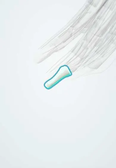
- Distal phalanx, middle finger
- Extensor tendons
- Distal phalanx, ring finger
- Distal phalanx, little finger
- Middle phalanx, middle finger
- Volar plates (fingers)
- Finger joint capsules
What it is
Loss of active extension of the fingertip at the distal interphalangeal (DIP) joint caused by disruption of the terminal extensor tendon, with or without a fracture of the base of the distal phalanx.
Who gets it
One of the most common tendon injuries of the hand. It is typical in ball sports, such as volleyball, baseball and basketball, and in everyday accidents like tucking in sheets. Middle, ring and little fingers are most often affected.
Causes
- Sudden forced flexion of the extended fingertip (ball hitting the tip)
- Jamming the finger against a hard surface
- Laceration or crush over the back of the DIP joint
- Hyperextension causing a bony avulsion
Risk factors
- Ball sports and catching
- Manual work
- Older age (minor trauma in degenerated tendons)
Pathophysiology
The terminal extensor tendon is the final part of the extensor mechanism and inserts on the dorsal base of the distal phalanx. When it ruptures or pulls off a bone fragment, the flexor digitorum profundus is unopposed and the tip droops. In chronic cases, the extensor force is concentrated at the PIP joint and the volar plate there can stretch, giving a swan-neck deformity.
Classification
- Tendinous (soft-tissue) mallet: tendon rupture without fracture
- Bony mallet: avulsion fracture of the dorsal base of the distal phalanx
- Doyle types I–IV (closed, open, deep abrasion, fracture types)
- Bony mallet with volar subluxation of the distal phalanx (unstable)
Symptoms and signs
- Drooping fingertip that cannot be actively straightened
- Full passive extension when the tip is lifted by the other hand
- Pain, swelling and bruising on the back of the DIP joint
- Swan-neck deformity in chronic untreated cases
Diagnosis
- Clinical
- Fingertip held flexed at the DIP joint with inability to extend it actively, while passive extension is preserved. The rest of the finger and the PIP joint are checked for associated injury.
- Tests
- Not needed.
- Imaging
- Lateral X-ray of the finger to identify a bony mallet, the size of the fragment and any volar subluxation of the distal phalanx.
Differential diagnosis
- Jersey finger (FDP avulsion: inability to flex the DIP joint)
- Distal phalanx fracture
- Central slip injury (boutonnière deformity)
- DIP joint dislocation
- Osteoarthritis of the DIP joint
Goal of treatment
Restore active extension of the fingertip by keeping the DIP joint continuously straight until the tendon or bone heals.
Treatment
- Conservative
- Continuous splinting of the DIP joint in extension (or slight hyperextension) for several weeks, leaving the PIP joint free; the tip must never drop during skin care or splint changes. Night splinting usually follows.
- Follow-up
- Follow-up X-rays in bony mallets to confirm the joint stays reduced in the splint.
- Surgical
- Surgery (pin fixation, extension block pinning or tendon repair) for open injuries, volar subluxation, large fragments, or patients who cannot keep a splint on.
- Rehabilitation
- Gradual flexion exercises after immobilisation.
Complications
- Persistent extensor lag (droop)
- Swan-neck deformity
- Skin breakdown under the splint
- DIP joint stiffness or arthritis
- Nail deformity or pin infection after surgery
Prognosis
Most closed mallet fingers do well with strict splinting, though a small residual lag is common. Results depend largely on adherence: if the tip drops during treatment, healing can be set back.
Test yourself
Think of the answer, then tap the question to see it.
Which tendon is disrupted in mallet finger, and where does it insert?
The terminal extensor tendon, which inserts on the dorsal base of the distal phalanx.
What is the standard treatment of a closed tendinous mallet finger?
Continuous splinting of the DIP joint in extension for several weeks, leaving the PIP joint free.
Which deformity can develop in an untreated chronic mallet finger?
Swan-neck deformity: DIP flexion with PIP hyperextension.
Which X-ray finding in a bony mallet usually points to surgical treatment?
Volar subluxation of the distal phalanx, often with a large fracture fragment.
Related conditions
Educational information, not medical advice. For any symptom, see a health professional.
Sources: Mallet Finger Injuries (StatPearls (NCBI Bookshelf), 2025).
Spotted a mistake?We are always improving for the community. Tell us what is wrong and our team will review it.Report a mistake
Representative model, for educational purposes only.