Conditions · Gastroenterology and hepatology
Mallory-Weiss syndrome !
A tear in the lining where the oesophagus meets the stomach, caused by forceful vomiting or retching, which makes a person vomit blood.
Updated 2026-11-01
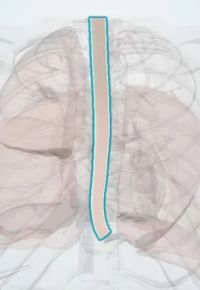
Structures involved
Tap one to see what it is and open it in 3D.
In the clinical exam
Why it happens
- Hemorrhage: petechiae, purpura, ecchymosis, hematoma, and what blood loss does
- Cachexia, anorexia nervosa and bulimia
- Alcohol: how it injures the liver, brain, heart and fetus
- Vomiting and its control
- Esophagus and the Barrett change
What it is
Upper gastrointestinal bleeding from a longitudinal mucosal laceration at the gastro-oesophageal junction or the cardia of the stomach, typically after forceful retching or vomiting. The tear involves mucosa and submucosa, not the full wall.
Who gets it
Accounts for a small share of upper GI bleeding episodes, mostly in adults aged 30 to 60 and more often in men. Heavy alcohol use is the classic association; in Colombia and Latin America binge drinking is a common trigger seen in emergency departments.
Causes
- Forceful retching or vomiting, classically after heavy alcohol intake
- Hyperemesis gravidarum and bulimia nervosa
- Severe coughing, straining, hiccups or blunt abdominal trauma
- Endoscopy or cardiopulmonary resuscitation (iatrogenic)
Risk factors
- Binge drinking or chronic alcohol use
- Hiatal hernia
- Conditions with repeated vomiting (gastroenteritis, chemotherapy, pregnancy)
- Portal hypertension or coagulopathy, which make bleeding heavier
Pathophysiology
During retching, a sudden rise in intra-abdominal pressure against a closed or poorly relaxing gastro-oesophageal junction distends the cardia. The shear force tears the mucosa longitudinally, usually on the lesser curve side, and exposes submucosal vessels, which bleed. Most tears stop bleeding spontaneously and heal within a few days.
Symptoms and signs
- Haematemesis (fresh blood or coffee-ground vomitus) after an episode of retching or non-bloody vomiting
- Epigastric or lower chest discomfort
- Melaena if bleeding is larger
- Dizziness or syncope with significant blood loss
Diagnosis
- Clinical
- Suspected when vomiting blood follows repeated retching; assess haemodynamic status first and estimate risk with a bleeding score such as Glasgow-Blatchford.
- Tests
- Complete blood count, coagulation tests, urea and creatinine, blood typing. Upper endoscopy confirms a longitudinal tear at the gastro-oesophageal junction and allows treatment if it is still bleeding.
- Imaging
- Chest radiograph or CT with oral contrast when perforation is suspected (pneumomediastinum, pleural effusion).
Differential diagnosis
- Boerhaave syndrome (full-thickness rupture)
- Oesophageal or gastric varices
- Peptic ulcer disease and erosive gastritis or oesophagitis
- Haemoptysis or swallowed blood from the nose
Goal of treatment
Stabilise the circulation, stop active bleeding and treat the cause of vomiting so the tear can heal.
Treatment
- Acute
- Intravenous access, fluid resuscitation and transfusion when needed; nil by mouth initially and antiemetics to stop retching.
- Medical
- Acid suppression with a proton pump inhibitor to help mucosal healing.
- Procedure
- Endoscopic haemostasis for active bleeding: clips, band ligation, thermal coagulation or epinephrine injection combined with another method; angiographic embolisation if endoscopy fails.
- Follow-up
- Nursing care: vital signs, amount and appearance of vomitus and stools, serial haemoglobin, aspiration precautions, and screening for alcohol use and withdrawal.
Complications
- Hypovolaemic shock
- Rebleeding
- Anaemia
- Progression to full-thickness perforation (rare)
Prognosis
Excellent in most cases: bleeding stops spontaneously in most patients and the tear heals within days. Death is uncommon and occurs mainly in patients with cirrhosis, coagulopathy or serious comorbidity.
Prevention
- Reducing harmful alcohol use
- Early treatment of persistent vomiting
Test yourself
Think of the answer, then tap the question to see it.
What is the classic history in Mallory-Weiss syndrome?
Haematemesis after repeated forceful retching or vomiting, often after an alcohol binge.
How deep is the tear, and how does that differ from Boerhaave syndrome?
Mallory-Weiss involves mucosa and submucosa only; Boerhaave is a full-thickness rupture with mediastinitis.
Where is the tear located?
Longitudinally at the gastro-oesophageal junction or proximal gastric cardia.
What is the usual course?
Most bleeding stops spontaneously; endoscopic therapy is used for active bleeding.
Related conditions
- Upper gastrointestinal bleeding
- Oesophageal perforation (Boerhaave syndrome)
- Hiatal hernia
- Alcohol use disorder
- Bulimia nervosa
- Peptic ulcer disease
- Portal hypertension
Educational information, not medical advice. For any symptom, see a health professional.
Sources: Mallory-Weiss tear: MedlinePlus Medical Encyclopedia (MedlinePlus (U.S. National Library of Medicine), 2024).
Spotted a mistake?We are always improving for the community. Tell us what is wrong and our team will review it.Report a mistake
Representative model, for educational purposes only.