Conditions · Gastroenterology and hepatology
Oesophageal perforation (Boerhaave syndrome) !
A full tear through the wall of the oesophagus, after forceful vomiting (Boerhaave) or a medical procedure. It is a surgical emergency.
Updated 2026-11-01
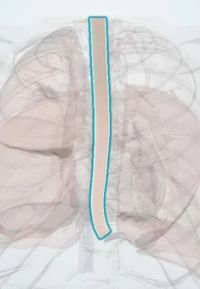
Structures involved
Tap one to see what it is and open it in 3D.
Why it happens
- Cachexia, anorexia nervosa and bulimia
- Vomiting and its control
- Swallowing and esophageal motility
- Esophagus and the Barrett change
What it is
A full-thickness disruption of the oesophageal wall that lets oesophageal and gastric contents leak into the mediastinum or pleural space. Boerhaave syndrome is the spontaneous form caused by a sudden rise in intraoesophageal pressure during vomiting; most perforations today are iatrogenic.
Who gets it
Uncommon but among the most lethal perforations of the gastrointestinal tract. Iatrogenic injury during endoscopy and dilation is the leading cause; Boerhaave syndrome typically affects middle-aged men after heavy eating or drinking.
Causes
- Iatrogenic: upper endoscopy, dilation, stent placement, nasogastric or transoesophageal echocardiography probes, thoracic surgery
- Spontaneous (Boerhaave): forceful vomiting against a closed upper oesophageal sphincter
- Foreign bodies, food impaction and caustic ingestion
- Penetrating or blunt trauma
- Underlying oesophageal disease: tumours, eosinophilic oesophagitis, strictures
Risk factors
- Therapeutic endoscopic procedures, especially dilation
- Binge eating and heavy alcohol intake with vomiting
- Oesophageal strictures, cancer or eosinophilic oesophagitis
- Older age and comorbidity (worse outcome)
Pathophysiology
In Boerhaave syndrome, vomiting raises pressure in the lower oesophagus while the upper sphincter stays closed, and the wall ruptures, usually in the left posterolateral distal oesophagus, just above the diaphragm, where the wall is weakest. Gastric contents and swallowed bacteria spill into the mediastinum, causing chemical and then bacterial mediastinitis, pleural effusion (usually left-sided), sepsis and multiorgan failure.
Classification
- By cause: iatrogenic, spontaneous (Boerhaave), traumatic, foreign body
- By site: cervical, thoracic or abdominal oesophagus
- By time to diagnosis: early (within 24 hours) or late, which strongly affects outcome
Symptoms and signs
- Severe retrosternal or upper abdominal pain after vomiting or a procedure
- Mackler triad: vomiting, lower chest pain and subcutaneous emphysema
- Dyspnoea, tachypnoea and pain on swallowing
- Fever, tachycardia and hypotension as mediastinitis and sepsis develop
- Neck pain and crepitus in cervical perforation
Diagnosis
- Clinical
- Suspect it in any patient with chest pain after vomiting or after instrumentation of the oesophagus; examine for crepitus in the neck and chest wall.
- Tests
- Blood counts, inflammatory markers, lactate, blood cultures. Pleural fluid from a thoracentesis may contain food particles, high amylase and low pH.
- Imaging
- Chest radiograph may show pneumomediastinum, left pleural effusion or hydropneumothorax. CT of the chest with oral water-soluble contrast is the test of choice to confirm the leak; a contrast oesophagram is an alternative.
Differential diagnosis
- Acute myocardial infarction
- Aortic dissection
- Pulmonary embolism and spontaneous pneumothorax
- Acute pancreatitis or perforated peptic ulcer
- Mallory-Weiss tear
Goal of treatment
Control the leak and the infection early, drain contaminated spaces, keep the patient nil by mouth with nutritional support, and restore oesophageal continuity.
Treatment
- Acute
- Resuscitation, nil by mouth, nasogastric decompression under guidance, early broad-spectrum intravenous antibiotics with antifungal cover when indicated, and acid suppression.
- Surgical
- Surgical repair of the perforation with debridement and drainage of the mediastinum and pleura, especially within 24 hours; resection or diversion when the oesophagus is diseased or the injury is extensive.
- Procedure
- Endoscopic stent or clip closure plus percutaneous drainage for selected contained or iatrogenic perforations; chest tube for pleural collections.
- Conservative
- Non-operative management only for small contained leaks in stable patients without sepsis, with close observation.
- Follow-up
- Intensive care: vital signs and sepsis signs, chest tube output, enteral (jejunal) or parenteral nutrition, and strict nil by mouth until healing is confirmed.
Complications
- Mediastinitis and empyema
- Sepsis and septic shock
- Multiorgan failure
- Oesophageal fistula or stricture
- Death
Prognosis
One of the most serious GI emergencies. Mortality is high and rises sharply when diagnosis and treatment are delayed beyond 24 hours; early treatment of an iatrogenic perforation carries a better outcome than spontaneous rupture.
Test yourself
Think of the answer, then tap the question to see it.
What is the Mackler triad?
Vomiting, lower chest pain and subcutaneous emphysema, classic for Boerhaave syndrome.
Where does a Boerhaave rupture usually occur?
Left posterolateral wall of the distal thoracic oesophagus, just above the diaphragm.
What is the most common cause of oesophageal perforation today?
Iatrogenic injury during endoscopy, especially dilation.
Which test best confirms the diagnosis?
CT of the chest with oral water-soluble contrast (or a water-soluble contrast oesophagram).
Why does timing matter so much?
Delay beyond 24 hours lets mediastinitis and sepsis develop and greatly increases mortality.
Related conditions
- Mallory-Weiss syndrome
- Sepsis and septic shock
- Pleural effusion
- Pneumothorax
- Myocardial Infarction (Heart Attack)
- Aortic dissection
- Acute pancreatitis
- Eosinophilic oesophagitis
- Oesophageal cancer
Educational information, not medical advice. For any symptom, see a health professional.
Sources: Esophageal perforation: MedlinePlus Medical Encyclopedia (MedlinePlus (U.S. National Library of Medicine), 2024).
Spotted a mistake?We are always improving for the community. Tell us what is wrong and our team will review it.Report a mistake
Representative model, for educational purposes only.