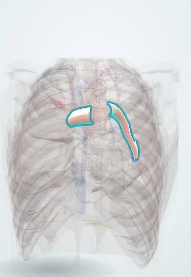
Pulmonary embolism !
A pulmonary embolism is a blockage of an artery in the lungs, usually by a blood clot that has travelled from a deep vein in the legs or pelvis.
Updated 2026-10-29
Structures involved
Tap one to see what it is and open it in 3D.
- Pulmonary arteries
- Pulmonary trunk
- Segmental lung vessels
- Right ventricle
- Right atrium
- Inferior vena cava
- Pleura
In biochemistry
In embryology
In the clinical exam
- Signs of deep vein thrombosis: Homans and the Wells score
- Shock: the clinical signs and the types
- Chest pain: ischemic, pericardial, pleural and other patterns
- Respiratory symptoms: cough, sputum, hemoptysis and dyspnea
In physiology
- Fibrinolysis and natural anticoagulants
- Thrombosis and bleeding: Virchow's triad and hemostatic defects
- The cardiac electrical axis
- Central venous pressure and the jugular pulse
- Dead space and alveolar ventilation
- The alveolar gas equation and the A–a gradient
- Ventilation–perfusion matching and West zones
- The causes of hypoxemia
- Diffusion across the respiratory membrane and DLCO
- The pulmonary circulation and hypoxic vasoconstriction
- Hypoxemic and hypercapnic respiratory failure
- The bronchial circulation
Why it happens
- Thrombosis and Virchow's triad
- Pulmonary thromboembolism: where it comes from and what it does
- What happens to a thrombus: propagation, embolism, dissolution, organization
- Infarction: red vs white, and what decides the damage
- The autopsy: clinical and medicolegal
- Shock: types, stages and the organs it damages
What it is
Blockage of a pulmonary artery or its branches, usually by a blood clot that has travelled from the deep veins of the legs or pelvis. It ranges from small, silent clots to sudden death.
Who gets it
A common cause of cardiovascular death and one of the most preventable causes of death in hospital. Pulmonary embolism and deep vein thrombosis are two forms of the same disease (venous thromboembolism).
Causes
- Thrombus from the deep veins of the legs or pelvis (most cases)
- Rarely fat, air, amniotic fluid or tumour emboli
Risk factors
- Recent surgery, especially orthopaedic, trauma or fracture
- Immobilisation, long travel, hospital admission
- Active cancer
- Pregnancy, postpartum, oestrogen-containing contraceptives or hormone therapy
- Previous venous thromboembolism or inherited thrombophilia
- Obesity, older age, heart failure
Pathophysiology
Virchow's triad (stasis, endothelial injury and hypercoagulability) favours a deep vein clot that breaks off and lodges in the pulmonary arteries. Blocked flow creates ventilation–perfusion mismatch and hypoxaemia, and raises pulmonary vascular resistance. The right ventricle dilates and can fail, lowering left-sided filling and causing shock.
Classification
- High-risk (massive): sustained hypotension or shock
- Intermediate-risk (submassive): normal blood pressure but right ventricular dysfunction and/or raised troponin
- Low-risk: no shock and no right ventricular strain (e.g., low PESI score)
- Acute vs chronic thromboembolic pulmonary hypertension
Symptoms and signs
- Sudden breathlessness (most common)
- Pleuritic chest pain
- Cough, sometimes haemoptysis
- Rapid breathing and fast heart rate
- Fainting
- Signs of deep vein thrombosis in a leg
Diagnosis
- Clinical
- Clinical probability with the Wells score or the revised Geneva score; in low-probability patients the PERC rule can exclude it without tests. Criteria and thresholds vary between guidelines.
- Tests
- D-dimer (age-adjusted in some protocols) to exclude it when probability is not high. Troponin and BNP for risk stratification. ECG: often sinus tachycardia; S1Q3T3 or right heart strain may appear. Arterial blood gas shows hypoxaemia.
- Imaging
- CT pulmonary angiography is the main test. Ventilation–perfusion scan when CT contrast is not suitable (e.g., pregnancy, kidney disease). Bedside echocardiography shows right ventricular dilation in unstable patients; compression ultrasound of the legs looks for DVT.
Differential diagnosis
- Acute coronary syndrome
- Pneumonia
- Pneumothorax
- Acute heart failure
- Aortic dissection
- Pericarditis or tamponade
Goal of treatment
Support breathing and circulation, stop clot growth and prevent recurrence; restore flow quickly in high-risk patients.
Treatment
- Acute
- Oxygen and haemodynamic support; immediate anticoagulation (heparins or direct oral anticoagulants) once the diagnosis is likely and bleeding risk is acceptable.
- Acute
- High-risk (massive) PE: systemic thrombolysis unless contraindicated.
- Procedure
- Catheter-directed therapy (thrombus aspiration or local thrombolysis) when thrombolysis fails or is contraindicated; vena cava filter only when anticoagulation is impossible.
- Surgical
- Surgical pulmonary embolectomy in selected high-risk patients.
- Medical
- Anticoagulation for at least about three months, longer if the cause persists or the event was unprovoked.
- Follow-up
- Close monitoring of intermediate-risk patients for decompensation; follow-up for chronic thromboembolic pulmonary hypertension.
Drugs for this condition
- Warfarin
- Heparin
- Rivaroxaban
- Dabigatran
- Apixaban
- Enoxaparin
- Fondaparinux
- Edoxaban
- Dalteparin
- Alteplase
- Streptokinase
Complications
- Sudden death and obstructive shock
- Right heart failure
- Pulmonary infarction and pleural effusion
- Recurrent venous thromboembolism
- Chronic thromboembolic pulmonary hypertension
- Bleeding from treatment
Prognosis
Most low-risk patients do well with anticoagulation, and some can be managed as outpatients. Mortality is high in massive PE, much of it in the first hours, which makes fast recognition essential.
Prevention
- Thromboprophylaxis in at-risk hospital and surgical patients
- Early mobilisation after surgery
- Mechanical prophylaxis when anticoagulants cannot be used
Test yourself
Think of the answer, then tap the question to see it.
Where do most pulmonary emboli come from?
From deep vein thrombosis in the legs or pelvis.
What is the main imaging test to confirm pulmonary embolism?
CT pulmonary angiography; a ventilation–perfusion scan is the alternative when contrast is not suitable.
What defines massive (high-risk) pulmonary embolism?
Pulmonary embolism with sustained hypotension, shock or cardiac arrest.
What is the role of the PERC rule?
In patients with low clinical probability, meeting all PERC criteria makes PE so unlikely that a D-dimer is not needed.
What is the reperfusion treatment of choice in massive PE without contraindications?
Systemic thrombolysis, with catheter-directed therapy or surgical embolectomy as alternatives.
Related conditions
- Deep vein thrombosis
- Myocardial Infarction (Heart Attack)
- Heart failure
- Pleural effusion
- Pneumothorax
- Aortic dissection
- Femoral shaft fracture
- Sepsis and septic shock
Educational information, not medical advice. For any symptom, see a health professional.
Sources: Acute Pulmonary Embolism (StatPearls (NCBI Bookshelf), 2024).
Spotted a mistake?We are always improving for the community. Tell us what is wrong and our team will review it.Report a mistake
Representative model, for educational purposes only.