
Radiation retinopathy
Months or years after radiotherapy near the eye, the small retinal vessels close and leak, which can slowly damage vision.
What it is
A delayed, slowly progressive occlusive retinal vasculopathy, usually 6 months to 3 years after radiation to the eye, orbit, sinuses or brain. It mimics diabetic retinopathy (microaneurysms, cotton wool spots, macular oedema, new vessels) and may come with radiation optic neuropathy.
Who gets it
Most common after plaque brachytherapy or proton beam therapy for uveal melanoma, where many patients develop it within a few years; also seen after radiotherapy for head and neck or skull base tumours.
Causes
- Plaque brachytherapy or proton beam therapy for intraocular tumours
- External beam radiotherapy for orbital, sinus, nasopharyngeal or brain tumours
Risk factors
- Higher total and per-fraction radiation dose to the retina
- Tumour close to the macula or optic disc
- Diabetes and hypertension
- Concurrent chemotherapy
Pathophysiology
Radiation kills capillary endothelial cells, which divide slowly, so damage appears late. Capillaries close, the retina becomes ischaemic and leaky, and VEGF drives macular oedema and new vessels.
Symptoms and signs
- Gradual painless loss of vision months to years after radiotherapy
- Floaters from vitreous haemorrhage
- Sometimes none in early stages
Diagnosis
- Clinical
- History of radiotherapy; fundoscopy shows microaneurysms, haemorrhages, cotton wool spots, hard exudates, capillary dropout and sometimes neovascularisation, confined to the irradiated field.
- Tests
- Blood glucose and blood pressure, since diabetes and hypertension worsen it.
- Imaging
- Fluorescein angiography shows capillary non-perfusion and leakage; OCT detects macular oedema; OCT angiography finds early capillary loss.
Differential diagnosis
- Diabetic retinopathy
- Hypertensive retinopathy
- Retinal vein occlusion
- Tumour recurrence
Microaneurysms and cotton wool spots: which vasculopathy?
| Feature | Radiation retinopathy | Diabetic retinopathy | Hypertensive retinopathy |
|---|---|---|---|
| History | Radiotherapy near the eye months to years before | Years of diabetes | High blood pressure, sometimes an acute crisis |
| Distribution | Confined to the irradiated field, may be one eye | Both eyes, fairly symmetrical | Both eyes |
| Typical signs | Capillary dropout, microaneurysms, cotton wool spots, sometimes optic neuropathy | Microaneurysms and dot-blot haemorrhages first, hard exudates | Narrowed arterioles, artery-vein nicking, flame haemorrhages, disc swelling if severe |
| Driver | Endothelial cell death after radiation | Chronic hyperglycaemia | Raised pressure damaging arterioles |
| Course | Often progresses with no further exposure | Slows with glucose and pressure control | Improves as pressure is controlled |
Goal of treatment
Control macular oedema and neovascularisation to preserve vision and the eye.
Treatment
- Medical
- Intravitreal anti-VEGF or corticosteroids for macular oedema, sometimes given early as prophylaxis after plaque therapy.
- Procedure
- Panretinal or sectoral laser photocoagulation of ischaemic retina when neovascularisation appears.
- Surgical
- Vitrectomy for non-clearing vitreous haemorrhage or traction.
- Follow-up
- Regular examination with OCT after any radiotherapy near the eye.
Complications
- Macular oedema and atrophy
- Vitreous haemorrhage
- Neovascular glaucoma
- Radiation optic neuropathy
- Radiation cataract
Common traps
- No retinopathy at the end of radiotherapy means the eye escaped: damage typically appears 6 months to 3 years later, so follow up with OCT for years
- New retinal changes after plaque therapy mean tumour recurrence: microaneurysms, cotton wool spots and capillary dropout in the field are vascular injury; recurrence shows as growth of the mass on ultrasound
- Diabetes and blood pressure do not matter once the cause is radiation: diabetes and hypertension damage the same small vessels and make radiation retinopathy worse
- Radiation retinopathy is untreatable: anti-VEGF or corticosteroid for oedema and laser for ischaemic retina preserve vision, and early treatment does better
- A red painful eye years after radiotherapy is surface irritation: high pressure with new vessels on the iris means neovascular glaucoma from ischaemic retina
Prognosis
Often progressive; vision depends on macular and optic nerve involvement. Early treatment of oedema improves outcomes.
Prevention
- Radiation planning that shields the macula and optic disc where possible
- Control of diabetes and blood pressure
Test yourself
Think of the answer, then tap the question to see it.
Why does radiation retinopathy appear months to years after treatment rather than during it?
Radiation kills cells mainly when they divide, and capillary endothelial cells divide slowly, so vessels close only as these cells turn over.
Two years after plaque brachytherapy for a choroidal melanoma, vision drops and OCT shows macular oedema. What is the likely cause and first treatment?
Radiation maculopathy. Intravitreal anti-VEGF or corticosteroid, plus laser to any ischaemic retina that produces new vessels.
Which examination finding points to radiation rather than diabetic retinopathy?
Changes confined to the irradiated field, often in one eye, after radiotherapy; diabetic retinopathy is bilateral and fairly symmetrical.
Which systemic conditions should be controlled to slow radiation retinopathy, and why?
Diabetes and hypertension, because they damage the same small vessels and add to the radiation injury.
Years after orbital radiotherapy, a patient has a painful red eye, high pressure and new vessels on the iris. What happened?
Neovascular glaucoma: ischaemic retina released VEGF, which drove new vessels into the drainage angle.
Why it happens
- Radiation oncology: principles for doctors
- Other retinal vascular disease: ROP and occlusions
- Ionizing radiation injury
Connections
Educational information, not medical advice. For any symptom, see a health professional.
Sources: Radiation Retinopathy (EyeWiki (American Academy of Ophthalmology), 2026).
Spotted a mistake?We are always improving for the community. Tell us what is wrong and our team will review it.Report a mistake
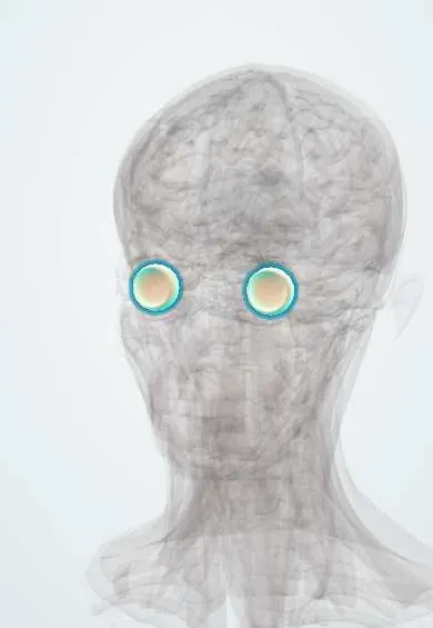
Representative model, for educational purposes only.