Conditions · Ophthalmology and ENT
Nasopharyngeal carcinoma !
A cancer of the upper part of the throat behind the nose, strongly linked to Epstein-Barr virus. A neck lump or fluid in one ear may be the first sign.
Updated 2026-11-01
Structures involved
Tap one to see what it is and open it in 3D.
- Pharynx
- Auditory tube
- Neck lymph nodes
- Nasal cavity
- Sphenoid sinus
- Abducens nerve (VI)
- Trigeminal nerve (V)
Microorganisms that cause it
Why it happens
- Chemical carcinogens: initiation, promotion and the table to know
- Diet, cancer and systemic disease
- Oncogenic viruses and H. pylori: the table to know
What it is
A malignant epithelial tumour of the nasopharynx, usually arising in the lateral recess (fossa of Rosenmüller) near the opening of the auditory tube. Most cases are the non-keratinising type associated with Epstein-Barr virus (EBV).
Who gets it
Endemic in southern China and Southeast Asia, and more common in North Africa and the Arctic; it is uncommon in Colombia and the rest of Latin America, where it is often diagnosed late. It peaks in middle age, with a smaller peak in adolescents, and is more frequent in men.
Causes
- Epstein-Barr virus infection, linked to most non-keratinising tumours
- Genetic susceptibility and family history
- Diet rich in salt-cured fish and preserved meats (nitrosamines), especially in early childhood
- Tobacco and heavy alcohol use, linked mainly to the keratinising type
Risk factors
- Asian, North African or Arctic ancestry
- Family history of nasopharyngeal carcinoma
- High antibody titres or plasma DNA of EBV
- Smoking and alcohol
- Occupational exposure to wood dust and formaldehyde
Pathophysiology
EBV latent proteins in infected epithelial cells drive proliferation and evade immunity. The tumour grows silently in the nasopharynx, blocks the auditory tube (causing serous otitis), spreads early to retropharyngeal and upper deep cervical lymph nodes, and can invade the skull base and cranial nerves, especially V and VI.
Classification
- Keratinising squamous cell carcinoma (WHO type I)
- Non-keratinising carcinoma, differentiated or undifferentiated (WHO types II and III, EBV-related, the endemic form)
- Basaloid squamous cell carcinoma
- TNM staging (AJCC)
Symptoms and signs
- Painless, firm lump in the upper neck (enlarged lymph node), often the first sign
- One-sided hearing loss, ear fullness or tinnitus from serous otitis media
- Nasal obstruction, blood-stained nasal discharge or nosebleeds
- Headache, facial numbness or double vision with skull base invasion
Diagnosis
- Clinical
- Nasal endoscopy (nasopharyngoscopy) to see the mass, full neck examination and cranial nerve examination.
- Tests
- Endoscopic biopsy confirms the diagnosis; EBV markers on the tissue; plasma EBV DNA is a tumour marker for screening in endemic areas, prognosis and follow-up. Fine-needle aspiration of a neck node that shows EBV-positive carcinoma points to a nasopharyngeal primary.
- Imaging
- MRI of the nasopharynx and neck defines local spread and skull base invasion; CT or PET-CT for staging and distant metastases (bone, lung, liver).
Differential diagnosis
- Adenoid hypertrophy (children)
- Lymphoma of the nasopharynx or neck
- Juvenile nasopharyngeal angiofibroma (adolescent boys with nosebleeds)
- Tuberculous cervical lymphadenitis
- Other head and neck squamous cell carcinomas with neck metastasis
Goal of treatment
Cure locoregional disease with high-precision radiotherapy, with chemotherapy for advanced stages, while protecting nearby structures and quality of life.
Treatment
- Procedure
- Radiotherapy, usually intensity-modulated, is the main treatment because the site is hard to reach surgically; early disease is treated with radiotherapy alone to the primary and both sides of the neck.
- Medical
- Locally advanced disease: concurrent chemoradiotherapy with platinum-based chemotherapy, often with induction or adjuvant chemotherapy; chemotherapy and immunotherapy (PD-1 inhibitors) for metastatic or recurrent disease.
- Surgical
- Neck dissection for persistent or recurrent neck nodes after radiotherapy; selected local recurrences may be resected.
- Follow-up
- Follow-up with endoscopy, MRI and plasma EBV DNA; nursing care for mucositis, dry mouth, nutrition, hearing, and thyroid function after neck irradiation.
Complications
- Skull base invasion and cranial nerve palsies
- Distant metastases to bone, lung and liver
- Radiotherapy effects: dry mouth, hearing loss, hypothyroidism, dental caries, trismus, neck fibrosis and osteoradionecrosis
- Persistent serous otitis media
Prognosis
Highly sensitive to radiotherapy; early-stage disease has high cure rates, while advanced and metastatic disease does worse. High plasma EBV DNA before or after treatment predicts a poorer outcome.
Prevention
- Avoiding tobacco and limiting salt-cured foods
- Plasma EBV DNA screening is used in some endemic populations
Test yourself
Think of the answer, then tap the question to see it.
Which virus is strongly associated with nasopharyngeal carcinoma?
Epstein-Barr virus.
Where in the nasopharynx does this cancer usually arise?
In the lateral recess, the fossa of Rosenmüller, near the auditory tube opening.
What ear finding in an adult should raise suspicion of this tumour?
Unilateral serous otitis media (middle ear effusion).
What is the main treatment modality?
Radiotherapy, combined with chemotherapy in locally advanced disease.
Related conditions
- Otitis media with effusion and chronic suppurative otitis media
- Infectious mononucleosis
- Cervical lymphadenopathy
- Head and neck squamous cell carcinoma (oral cavity and oropharynx, HPV)
- Hodgkin lymphoma
- Non-Hodgkin lymphoma
- Epistaxis (nosebleed)
Educational information, not medical advice. For any symptom, see a health professional.
Sources: Nasopharyngeal Carcinoma Treatment (PDQ®)–Health Professional Version (National Cancer Institute (NCI), 2026); Nasopharyngeal Cancer Treatment (PDQ®)–Patient Version (National Cancer Institute (NCI), 2026).
Spotted a mistake?We are always improving for the community. Tell us what is wrong and our team will review it.Report a mistake
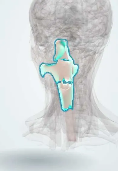
Representative model, for educational purposes only.