Conditions · Hematology and oncology
Soft tissue sarcomas (liposarcoma, leiomyosarcoma, rhabdomyosarcoma) !
Rare cancers of fat, muscle and other connective tissue; most often a painless lump deep in the thigh that keeps growing.
Updated 2026-11-01
Structures involved
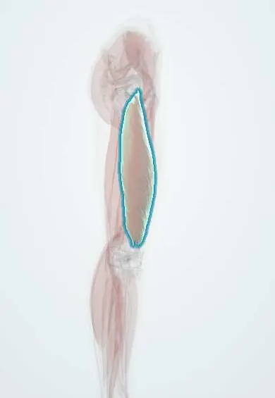
Tap one to see what it is and open it in 3D.
Why it happens
- Differentiation and anaplasia: how a tumour cell looks under the microscope
- Pathways of spread and the usual metastatic sites
- Naming tumours: -oma, carcinoma, sarcoma, and the misleading names
- Ionizing radiation injury
- The cell cycle in cancer: cyclins, CDKs and checkpoints
- Grading and staging: what TNM means
Show 4 more
What it is
A group of more than 50 malignant tumours that arise from mesenchymal tissue such as fat (liposarcoma), smooth muscle (leiomyosarcoma), skeletal muscle (rhabdomyosarcoma), fibrous tissue and nerve sheaths. They usually present as a deep mass in a limb, most often the thigh, and spread mainly through the blood to the lungs.
Who gets it
Rare, under 1% of adult cancers. About 45% arise in the limbs, many of the rest in the abdomen and retroperitoneum, the trunk and the head and neck. Liposarcoma and undifferentiated pleomorphic sarcoma are frequent in adults, leiomyosarcoma in the uterus and retroperitoneum, and rhabdomyosarcoma is the commonest soft tissue sarcoma of children. In Colombia, delays in referral of a 'lump' to a sarcoma centre are a recognised problem.
Causes
- Most cases arise without a known cause
- Previous radiotherapy to the area, often years earlier
- Inherited syndromes: Li–Fraumeni (TP53), neurofibromatosis type 1 (malignant nerve sheath tumour) and hereditary retinoblastoma (RB1)
- Chronic lymphoedema (lymphangiosarcoma, for example after breast cancer surgery)
- Chemical exposures such as vinyl chloride, thorium dioxide and arsenic (hepatic angiosarcoma)
Risk factors
- Prior radiation therapy
- Family history suggesting Li–Fraumeni syndrome
- Neurofibromatosis type 1
- Long-standing lymphoedema
- Childhood (rhabdomyosarcoma) or older age (most adult types)
Pathophysiology
Malignant mesenchymal cells grow along fascial planes, pushing aside muscle and forming a pseudocapsule that contains microscopic tumour extensions, which is why shelling out the lump causes local recurrence. Many subtypes carry characteristic chromosomal translocations (for example in synovial sarcoma, myxoid liposarcoma and alveolar rhabdomyosarcoma) or gene amplifications such as MDM2 in well-differentiated and dedifferentiated liposarcoma. Spread is mainly haematogenous to the lungs; lymph node spread is uncommon except in a few subtypes.
Classification
- By tissue of origin: liposarcoma, leiomyosarcoma, rhabdomyosarcoma (embryonal, alveolar, pleomorphic), synovial sarcoma, undifferentiated pleomorphic sarcoma, malignant peripheral nerve sheath tumour and others
- Histological grade 1–3 (differentiation, mitotic count and necrosis)
- TNM staging by size, depth, grade, nodes and metastases
- By site: extremity and trunk, retroperitoneal, visceral, head and neck
Symptoms and signs
- Painless, enlarging lump, often deep to the fascia
- Pain or swelling only when the mass presses on nerves or vessels
- Abdominal fullness or vague pain with retroperitoneal tumours, which can grow very large
- In children with rhabdomyosarcoma: a mass in the head and neck, orbit or genitourinary tract
- Cough or breathlessness only with lung metastases
Diagnosis
- Clinical
- Every deep or large or growing soft tissue lump is treated as a possible sarcoma until proved otherwise and referred to a sarcoma team before any surgery. An unplanned excision ('whoops' operation) contaminates tissue planes and worsens outcomes.
- Tests
- Image-guided core needle biopsy, planned so the tract can be removed at surgery, with review by an expert pathologist using immunohistochemistry and molecular tests (for example MDM2 amplification or specific translocations). Grade is set on the biopsy.
- Imaging
- MRI of the primary site to define size, depth and relation to nerves and vessels; CT of the chest to look for lung metastases; CT of the abdomen and pelvis for retroperitoneal tumours; PET-CT in selected cases.
Differential diagnosis
- Lipoma and other benign soft tissue tumours
- Haematoma, especially in a patient taking anticoagulants
- Abscess
- Bone sarcoma with soft tissue extension (osteosarcoma, Ewing sarcoma)
- Lymphoma or metastatic carcinoma in soft tissue
- Myositis ossificans
Goal of treatment
Remove the tumour completely with a wide margin while keeping the limb and its function, and reduce the risk of local and distant recurrence.
Treatment
- Surgical
- Wide excision with a cuff of normal tissue, preserving the limb in most cases; amputation is reserved for tumours that cannot otherwise be removed.
- Procedure
- Radiotherapy before or after surgery for high-grade, large or deep tumours, or when margins are close, to reduce local recurrence.
- Medical
- Chemotherapy (anthracycline-based, sometimes with ifosfamide) for advanced or metastatic disease and selected high-risk tumours; multi-agent chemotherapy is standard for rhabdomyosarcoma in children. Some subtypes respond to targeted drugs.
- Follow-up
- Follow-up with clinical examination, imaging of the operated site and chest imaging, most intense in the first few years; rehabilitation for limb function.
Complications
- Local recurrence, more likely after unplanned or marginal excision
- Lung metastases
- Wound complications and fibrosis after radiotherapy and surgery
- Loss of limb function or amputation
- Cardiotoxicity from anthracyclines
Prognosis
Small, superficial, low-grade tumours are often cured by surgery alone. Large, deep, high-grade tumours, age over 60 and positive margins carry a higher risk of lung metastases and death; resection of a few lung metastases can still give long survival in selected patients.
Test yourself
Think of the answer, then tap the question to see it.
What is the commonest site of soft tissue sarcoma in adults?
The limbs, especially the thigh.
Where do soft tissue sarcomas most often metastasise?
To the lungs, through the bloodstream.
How should a deep soft tissue mass larger than 5 cm be investigated?
MRI and an image-guided core biopsy planned by a sarcoma team before any excision.
Which is the most common soft tissue sarcoma in children?
Rhabdomyosarcoma.
Which inherited syndromes predispose to soft tissue sarcomas?
Li–Fraumeni syndrome (TP53) and neurofibromatosis type 1, among others.
Related conditions
- Osteosarcoma
- Ewing sarcoma
- Kaposi sarcoma
- Neurofibromatosis (types 1 and 2)
- Gastrointestinal stromal tumour (GIST)
- Lymphoedema
- Retinoblastoma
- Lung cancer
Educational information, not medical advice. For any symptom, see a health professional.
Sources: Soft Tissue Sarcoma Treatment (PDQ®)–Health Professional Version (National Cancer Institute, 2025).
Spotted a mistake?We are always improving for the community. Tell us what is wrong and our team will review it.Report a mistake
Representative model, for educational purposes only.