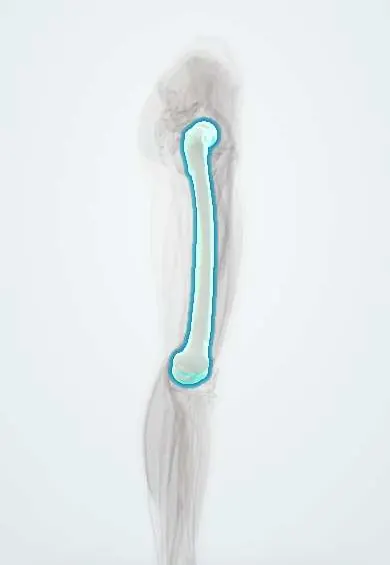
Conditions · Surgery and trauma
Osteosarcoma !
Osteosarcoma is the most common primary bone cancer in teenagers. It grows near the knee or shoulder, causes pain and swelling, and often spreads to the lungs.
Updated 2026-11-01
Structures involved
Tap one to see what it is and open it in 3D.
Why it happens
- Tumour suppressor genes: RB, TP53, APC, BRCA and the two-hit hypothesis
- Pathways of spread and the usual metastatic sites
- Tumours of infancy and childhood
- Invasion and metastasis: the metastatic cascade, step by step
- Radiation and cancer: ultraviolet and ionizing
- Bone tissue: cells, compact and spongy bone
What it is
A malignant primary bone tumour in which the cancer cells produce osteoid (immature bone). It usually arises in the metaphysis of long bones during the adolescent growth spurt, most often the distal femur, proximal tibia and proximal humerus.
Who gets it
The most common primary malignant bone tumour in children and adolescents, with a main peak in the second decade and a second peak in older adults, often secondary to Paget disease of bone or prior radiation. In Colombia and Latin America many patients still present with large tumours or lung metastases, which affects survival.
Causes
- Mostly sporadic, linked to rapid bone growth
- Inherited syndromes: hereditary retinoblastoma (RB1), Li-Fraumeni syndrome (TP53), Rothmund-Thomson syndrome
- Prior radiation therapy
- Paget disease of bone in older adults
Risk factors
- Adolescence and the growth spurt
- Tall stature and male sex
- Hereditary retinoblastoma or Li-Fraumeni syndrome
- Previous radiotherapy or chemotherapy
- Paget disease of bone
Pathophysiology
Malignant mesenchymal cells, often with RB1 and TP53 pathway alterations, form disorganised osteoid. The tumour destroys the cortex, lifts the periosteum and invades soft tissue. It spreads through the blood, most often to the lungs and then other bones; micrometastases are presumed present at diagnosis, which is why systemic chemotherapy is needed.
Classification
- Conventional high-grade (osteoblastic, chondroblastic, fibroblastic)
- Telangiectatic osteosarcoma
- Surface osteosarcomas: parosteal (low grade) and periosteal
- Secondary osteosarcoma (after radiation or Paget disease)
- Localised versus metastatic at diagnosis
Symptoms and signs
- Bone pain near the knee or shoulder, often worse at night and with activity
- Swelling or a firm mass
- Limp or reduced joint movement
- Pathological fracture after minor trauma
- Pain often first attributed to a sports injury or growing pains
Diagnosis
- Clinical
- Suspected in an adolescent with persistent pain and swelling around the knee or shoulder. The biopsy is planned with the treating sarcoma team so the tract can be removed at surgery.
- Tests
- Core-needle or open biopsy confirms the diagnosis; alkaline phosphatase and lactate dehydrogenase are often raised and carry prognostic value.
- Imaging
- Plain X-ray shows a destructive metaphyseal lesion with mixed lytic and sclerotic areas, a sunburst pattern and periosteal elevation (Codman triangle). MRI of the whole bone defines local extent and skip lesions; chest CT and bone scan or PET stage the disease.
Differential diagnosis
- Ewing sarcoma (diaphysis, onion-skin periosteal reaction)
- Osteomyelitis
- Benign bone tumours such as aneurysmal bone cyst or giant cell tumour
- Stress fracture or myositis ossificans
- Chondrosarcoma in older adults
Goal of treatment
Cure through control of both the primary tumour and micrometastatic disease while preserving limb function where possible.
Treatment
- Medical
- Multi-agent chemotherapy before surgery (neoadjuvant) and after it (adjuvant); the degree of tumour necrosis after preoperative chemotherapy guides prognosis.
- Surgical
- Wide resection with clear margins: limb-salvage surgery with endoprosthesis or bone graft in most patients, and amputation or rotationplasty when salvage is not safe. Resectable lung metastases are removed surgically.
- Procedure
- Radiotherapy is reserved for unresectable sites or positive margins because osteosarcoma is relatively radioresistant.
- Rehabilitation
- Physiotherapy and prosthetic rehabilitation after limb salvage or amputation, and long-term follow-up for late effects.
Complications
- Lung metastases
- Pathological fracture
- Local recurrence
- Chemotherapy toxicity: hearing loss, heart and kidney damage, infertility
- Prosthesis failure or infection after limb salvage
Prognosis
Long-term survival is about 70% for localised disease treated with chemotherapy and surgery, but much lower when metastases are present at diagnosis. Good response to preoperative chemotherapy and complete resection improve outcomes.
Test yourself
Think of the answer, then tap the question to see it.
What are the most common sites of osteosarcoma?
The metaphyses around the knee (distal femur, proximal tibia) and the proximal humerus.
Which X-ray signs are classic for osteosarcoma?
A sunburst periosteal reaction and a Codman triangle from periosteal elevation.
Which inherited conditions predispose to osteosarcoma?
Hereditary retinoblastoma (RB1) and Li-Fraumeni syndrome (TP53).
What is the most common site of osteosarcoma metastasis?
The lungs.
Which condition predisposes older adults to secondary osteosarcoma?
Paget disease of bone, as well as prior radiation therapy.
Related conditions
Educational information, not medical advice. For any symptom, see a health professional.
Sources: Osteosarcoma and Undifferentiated Pleomorphic Sarcoma of Bone Treatment (PDQ®)–Health Professional Version (National Cancer Institute, 2025); Osteosarcoma: MedlinePlus Medical Encyclopedia (MedlinePlus (U.S. National Library of Medicine), 2024).
Spotted a mistake?We are always improving for the community. Tell us what is wrong and our team will review it.Report a mistake
Representative model, for educational purposes only.