Conditions · Hematology and oncology
Gastrointestinal stromal tumour (GIST) !
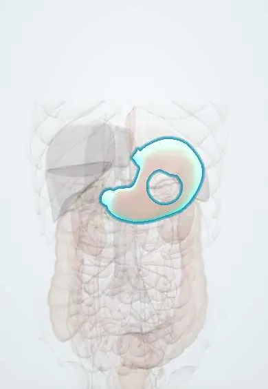
The most common sarcoma of the gut, usually in the stomach. It can bleed or grow silently, and most respond to a targeted pill called imatinib.
Updated 2026-11-01
Structures involved
Tap one to see what it is and open it in 3D.
Why it happens
- Oncogenes: RAS, MYC, HER2, BCR-ABL and the table to know
- Immunohistochemistry: the principle and the markers to know
- Molecular pathology: PCR, FISH, sequencing and liquid biopsy
- Tumour markers: serum and tissue, and what each one is for
- Receptor tyrosine kinases
- The GI tract wall: mucosa, submucosa, muscularis, serosa; enteric plexuses
What it is
A mesenchymal tumour of the digestive tract that arises from the interstitial cells of Cajal, the pacemaker cells of gut motility. Most are driven by activating mutations in KIT or PDGFRA, and their behaviour ranges from tiny harmless nodules to aggressive cancers that spread to the liver and peritoneum.
Who gets it
The most common mesenchymal tumour of the gut, though under 1% of digestive cancers. It occurs mainly in adults aged 50–70, equally in men and women. About 60% arise in the stomach and 30% in the small intestine, with the rest in the duodenum, rectum, colon or oesophagus. Small GISTs are often found by chance during endoscopy or surgery.
Causes
- Activating KIT mutations (about 80%)
- PDGFRA mutations (about 5–10%), including D842V, which resists imatinib
- KIT/PDGFRA wild-type tumours: SDH-deficient GIST (children and young adults, Carney triad and Carney–Stratakis syndrome), NF1-associated GIST, BRAF-mutant
- Rare familial germline KIT or PDGFRA mutations
Risk factors
- Age 50–70
- Neurofibromatosis type 1 (often multiple small-bowel GISTs)
- Succinate dehydrogenase deficiency syndromes
- No known environmental or dietary risk factors
Pathophysiology
KIT and PDGFRA are tyrosine kinase receptors that normally switch on only when their growth factor binds. Mutations keep them switched on, sending constant growth and survival signals to Cajal-cell precursors. The tumour grows outward from the muscle wall, so it often stays silent until it is large; it can ulcerate the mucosa and bleed, block the lumen or rupture into the abdomen. Spread is mainly to the liver and peritoneum; unlike carcinomas, lymph node spread is rare.
Classification
- Risk of recurrence by tumour size, mitotic rate and site (gastric tumours are less aggressive than small-bowel or rectal ones)
- Tumour rupture as a very high-risk feature
- Molecular subtype: KIT exon 11 or 9, PDGFRA, SDH-deficient, NF1-associated
- Localised, locally advanced or metastatic
Symptoms and signs
- Often none; found incidentally
- Gastrointestinal bleeding: melaena, vomiting blood or chronic occult bleeding with anaemia
- Abdominal pain, fullness or early satiety
- Palpable abdominal mass
- Bowel obstruction or difficulty swallowing depending on site
Diagnosis
- Clinical
- Suspect it in a patient with gut bleeding or anaemia and a submucosal mass on endoscopy, or an intra-abdominal mass arising from the bowel wall on imaging.
- Tests
- Endoscopy shows a smooth submucosal bulge, sometimes with central ulceration; endoscopic ultrasound-guided biopsy (avoiding percutaneous biopsy that may seed the peritoneum). Histology shows spindle or epithelioid cells; immunohistochemistry is positive for CD117 (KIT) and DOG1. Mutation analysis of KIT and PDGFRA guides drug choice. Complete blood count for anaemia.
- Imaging
- Contrast CT of the abdomen and pelvis for staging and follow-up; MRI for rectal and liver lesions; FDG-PET can show early response to imatinib.
Differential diagnosis
- Leiomyoma and leiomyosarcoma
- Schwannoma
- Gastric adenocarcinoma
- Gastrointestinal lymphoma
- Neuroendocrine tumour
- Ectopic pancreas or other submucosal lesions
Goal of treatment
Remove localised tumours completely without rupture, lower recurrence in high-risk cases with targeted therapy, and control metastatic disease with tyrosine kinase inhibitors chosen by mutation.
Treatment
- Surgical
- Complete resection with an intact capsule and negative margins, often laparoscopic; routine lymph node dissection is not needed because nodal spread is rare. Very small gastric tumours may be followed endoscopically.
- Medical
- Imatinib, a tyrosine kinase inhibitor, is given for at least three years after surgery in high-risk tumours, before surgery to shrink large ones, and as first-line treatment for metastatic disease; avapritinib is used for PDGFRA D842V tumours.
- Medical
- After imatinib resistance: sunitinib, then regorafenib and ripretinib. Conventional chemotherapy and radiotherapy are largely ineffective.
- Follow-up
- CT follow-up for recurrence; nursing and pharmacy support for adherence and side effects of imatinib such as oedema, diarrhoea, nausea, fatigue and low blood counts, and for drug interactions.
Complications
- Acute or chronic gastrointestinal bleeding
- Tumour rupture with intra-abdominal haemorrhage and peritoneal seeding
- Bowel obstruction
- Liver and peritoneal metastases
- Resistance to tyrosine kinase inhibitors
Prognosis
Small, low-mitotic gastric GISTs are usually cured by surgery. High-risk and metastatic tumours relapse often, but targeted therapy has turned metastatic GIST from a disease with survival of about a year and a half into one where many patients live five years or more.
Test yourself
Think of the answer, then tap the question to see it.
From which cells do GISTs arise?
The interstitial cells of Cajal, the pacemaker cells of the gut.
Which two immunostains are typically positive in GIST?
CD117 (KIT) and DOG1.
What is the first-line drug for metastatic KIT-mutant GIST?
Imatinib, a tyrosine kinase inhibitor.
Why is routine lymphadenectomy not done in GIST surgery?
Because GISTs rarely spread to lymph nodes; they spread to liver and peritoneum.
What is the most common site of GIST?
The stomach (about 60%), followed by the small intestine.
Related conditions
- Gastric cancer
- Upper gastrointestinal bleeding
- Lower gastrointestinal bleeding
- Iron deficiency anaemia
- Small bowel obstruction
- Peptic ulcer disease
- Neurofibromatosis (types 1 and 2)
- Soft tissue sarcomas (liposarcoma, leiomyosarcoma, rhabdomyosarcoma)
- Chronic myeloid leukaemia
Educational information, not medical advice. For any symptom, see a health professional.
Sources: Gastrointestinal Stromal Tumors Treatment (PDQ®)–Health Professional Version (National Cancer Institute, 2025).
Spotted a mistake?We are always improving for the community. Tell us what is wrong and our team will review it.Report a mistake
Representative model, for educational purposes only.