Conditions · Surgery and trauma
Ewing sarcoma !
Ewing sarcoma is a cancer of bone or nearby soft tissue in children and teenagers. It often starts in the pelvis or leg bones and is treated with chemotherapy.
Updated 2026-11-01
Structures involved
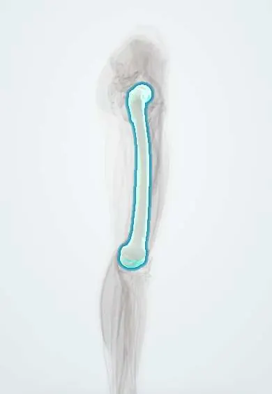
Tap one to see what it is and open it in 3D.
Why it happens
- Tumours of infancy and childhood
- Translocations, amplifications and epigenetic changes in cancer
- Molecular pathology: PCR, FISH, sequencing and liquid biopsy
- Immunohistochemistry: the principle and the markers to know
What it is
A malignant small round blue cell tumour of bone, and less often of soft tissue, defined by a fusion of the EWSR1 gene with an ETS-family transcription factor gene. It is the second most common primary bone cancer of childhood after osteosarcoma and classically involves the diaphysis of long bones and the flat bones of the pelvis and chest wall.
Who gets it
Rare: a few cases per million people under 20 each year, peaking in adolescence (10–20 years) and slightly more frequent in males. It is much more common in people of European ancestry than in people of African ancestry. In Colombia and Latin America, bone sarcomas in children are treated in paediatric oncology referral centres, and delayed diagnosis is common because pain is first attributed to sports injuries or growing pains.
Causes
- Somatic chromosomal translocation t(11;22)(q24;q12) creating the EWSR1::FLI1 fusion in most cases
- Less common fusions such as EWSR1::ERG
- Secondary changes such as trisomy 8 and loss of tumour-suppressor genes
- No proven environmental cause; it is not linked to radiation or to an inherited cancer syndrome in most patients
Risk factors
- Age 10–20 years
- Male sex
- European ancestry
- No modifiable risk factor is known; most patients have none
Pathophysiology
The fusion protein acts as an abnormal transcription factor that switches on growth genes and blocks differentiation, producing sheets of uniform small round cells (CD99 positive) that may form pseudorosettes. The tumour grows from the marrow cavity through the cortex, lifting the periosteum in layers and forming a large soft-tissue mass. It spreads early by blood to the lungs, other bones and the bone marrow, and can cause fever and raised inflammatory markers that mimic infection.
Classification
- Localised disease versus metastatic disease at diagnosis (the strongest prognostic factor)
- Skeletal primary (pelvis, femur, tibia/fibula, ribs, spine) versus extraskeletal soft-tissue primary
- Ewing family of tumours includes the former peripheral primitive neuroectodermal tumour (PNET) and Askin tumour of the chest wall
- Related undifferentiated small round cell sarcomas without EWSR1–ETS fusion (CIC- or BCOR-rearranged) are now classified separately
Symptoms and signs
- Persistent, often night-time bone pain that worsens over weeks
- Swelling or a palpable, sometimes warm and tender mass
- Fever, weight loss and fatigue
- Limp or reduced movement of the limb
- Pathological fracture after minor trauma
Diagnosis
- Clinical
- History of progressive bone pain and swelling in an adolescent, often with systemic symptoms. Any suspected primary bone tumour should be referred to a sarcoma centre before biopsy, so the biopsy track can later be removed with the tumour.
- Tests
- Core needle or open biopsy planned by the treating surgeon, with histology (small round blue cells, CD99 membrane staining) and molecular confirmation of the EWSR1 fusion. Full blood count, LDH (high values carry a worse prognosis) and bone marrow evaluation for staging.
- Imaging
- Plain radiograph: permeative, moth-eaten lytic lesion in the diaphysis with layered periosteal reaction (onion skin) and a soft-tissue mass. MRI of the whole bone defines local extent; CT of the chest looks for lung metastases; PET-CT or bone scan stages the skeleton.
Differential diagnosis
- Osteomyelitis (fever, raised markers and a lytic lesion)
- Osteosarcoma (metaphyseal, bone-forming tumour with sunburst periosteal reaction)
- Langerhans cell histiocytosis
- Lymphoma or leukaemia involving bone
- Metastatic neuroblastoma in younger children
Goal of treatment
Cure the disease by controlling micrometastases with systemic therapy and removing or irradiating the primary tumour, while preserving the limb and function where possible.
Treatment
- Medical
- Multi-agent chemotherapy is given to every patient, before local treatment (neoadjuvant) and after it, because most patients have undetectable micrometastases at diagnosis.
- Surgical
- Wide surgical resection of the primary tumour, usually with limb-sparing reconstruction; amputation is reserved for selected cases.
- Procedure
- Radiotherapy, since Ewing sarcoma is radiosensitive: used alone where resection is not feasible (for example some pelvic and spinal tumours), after incomplete resection, and for metastases.
- Medical
- High-dose chemotherapy with autologous stem cell rescue in selected high-risk patients, within clinical trials or specialised protocols.
- Follow-up
- Nursing priorities: neutropenic fever precautions, central line care, nausea and nutrition support, fracture precautions on the affected bone, pain control, psychosocial support for the family and school.
- Rehabilitation
- Physiotherapy and prosthetic or orthotic rehabilitation after limb surgery, with long-term survivorship follow-up.
Complications
- Lung, bone and bone marrow metastases
- Pathological fracture
- Spinal cord compression with spinal or paraspinal tumours
- Chemotherapy toxicity: neutropenic sepsis, cardiotoxicity, infertility and secondary leukaemia
- Radiation-associated growth disturbance and second sarcomas
- Local recurrence
Prognosis
About two thirds or more of patients with localised disease are cured with combined treatment. Metastatic disease at diagnosis lowers survival considerably, and isolated lung metastases do better than bone or marrow spread. Large tumours, pelvic primaries, high LDH and poor response to initial chemotherapy also worsen the outlook.
Test yourself
Think of the answer, then tap the question to see it.
Which chromosomal translocation is characteristic of Ewing sarcoma?
t(11;22)(q24;q12), producing the EWSR1::FLI1 fusion gene.
What periosteal reaction is classically described on radiographs of Ewing sarcoma?
A layered 'onion skin' periosteal reaction around a permeative diaphyseal lytic lesion.
Which immunohistochemical marker is strongly expressed on the membrane of Ewing sarcoma cells?
CD99.
Which part of a long bone does Ewing sarcoma typically involve, compared with osteosarcoma?
Ewing sarcoma favours the diaphysis; osteosarcoma favours the metaphysis.
What is the most important prognostic factor in Ewing sarcoma?
Whether metastatic disease is present at diagnosis.
Related conditions
- Osteosarcoma
- Osteomyelitis
- Langerhans cell histiocytosis
- Neuroblastoma
- Acute leukaemia (AML and ALL)
- Non-Hodgkin lymphoma
Educational information, not medical advice. For any symptom, see a health professional.
Sources: Ewing Sarcoma and Undifferentiated Small Round Cell Sarcomas of Bone and Soft Tissue Treatment (PDQ®)–Health Professional Version (National Cancer Institute, 2025); Ewing sarcoma: MedlinePlus Medical Encyclopedia (MedlinePlus (U.S. National Library of Medicine), 2025).
Spotted a mistake?We are always improving for the community. Tell us what is wrong and our team will review it.Report a mistake
Representative model, for educational purposes only.