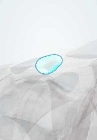
Joints and ligaments · Upper limb
AC capsule !
The acromioclavicular capsule seals the joint between the collarbone and the tip of the shoulder blade; it tears in a shoulder separation.
Official name: Acromioclavicular joint capsule
Opens in the Durgical app, no sign-up needed.
What it is
The capsule of the joint at the top of the shoulder.
What it does
Seals the AC joint; it is torn in a shoulder separation.
Key facts
- Attaches
- Margins of the acromion and the lateral end of the clavicle
- Function
- Thin; reinforced above by the acromioclavicular ligament and the deltoid-trapezius fascia
Relations
- Above
- Deltoid and trapezius aponeuroses blend over it; the skin is thin, so the clavicle end is easy to feel
- Below
- Subacromial bursa and supraspinatus tendon
- Medial
- Conoid ligament and trapezoid ligament, a little medial, running from the clavicle's underside to the coracoid
- Contents
- AC disc, often incomplete and wearing away with age
How to find it
The lateral end of the clavicle is followed outward until a small step or dip marks the joint, just medial to the edge of the acromion; bringing the arm across the chest moves the joint under the finger.
Examination
- Cross-body adduction (scarf) test: pain over the joint when the arm is brought across the chest suggests AC joint disease
- O'Brien active compression test: pain on top of the shoulder (rather than deep inside) points to the AC joint
- Pain relief after local anaesthetic is placed in the joint supports it as the source
On imaging
The Zanca view (AP with the beam tilted about 10–15° upward) shows the joint clear of the scapular spine. Comparison with the other side and the coracoclavicular distance grade a separation; MRI shows capsular oedema and cysts.
Clinical notes
- Distal clavicle osteolysis in weightlifters: pain and bone resorption at the clavicle end
- Distal clavicle excision for a painful arthritic joint keeps the superior and posterior capsule to avoid instability
- Type III separations are often managed without surgery; types IV–VI are usually reduced and reconstructed
Anatomical variants
- A coracoclavicular joint or bony bar between the clavicle and the coracoid is an occasional finding (close to 1 in 10 in one large series)
Development
The clavicle ossifies mainly in membrane and the acromion from cartilage; tissue left between them may persist as an articular disc, which is often missing or only partial and wears down early in adult life.
Related conditions
Test yourself
Think of the answer, then tap the question to see it.
What does the AC capsule with the acromioclavicular ligament resist, compared with the coracoclavicular ligaments?
Front-to-back movement of the clavicle; the coracoclavicular ligaments resist upward displacement.
What structures are torn in a Rockwood type III AC separation?
The AC capsule and ligament plus the coracoclavicular (conoid and trapezoid) ligaments.
Which test suggests AC joint disease?
The cross-body adduction test: pain over the AC joint when the arm is brought across the chest.
Educational information, not medical advice. For any symptom, see a health professional.
Its names
- Official Latin (TA2)
- capsula articularis articulationis acromioclavicularis TA2 1536
- Official Spanish (DeCS)
- Cápsula articular de la articulación acromioclavicular
- English (TA2)
- Acromioclavicular joint capsule
Related
Sources: Terminologia Anatomica (TA2); Anatomy, Shoulder and Upper Limb, Acromioclavicular Joint; 8.5 Development of the Appendicular Skeleton (OpenStax (Rice University), 2022) · adapted, CC BY 4.0; Anatomy of the Human Body (20th ed.): Development of the Joints (Lea & Febiger (Gray's Anatomy, 1918), 1918) · adapted, Public domain; Anatomy of the Human Body (20th ed.): Acromioclavicular Articulation (Lea & Febiger (Gray's Anatomy, 1918), 1918) · adapted, Public domain.
Spotted a mistake?We are always improving for the community. Tell us what is wrong and our team will review it.Report a mistake
Representative model, for educational purposes only.