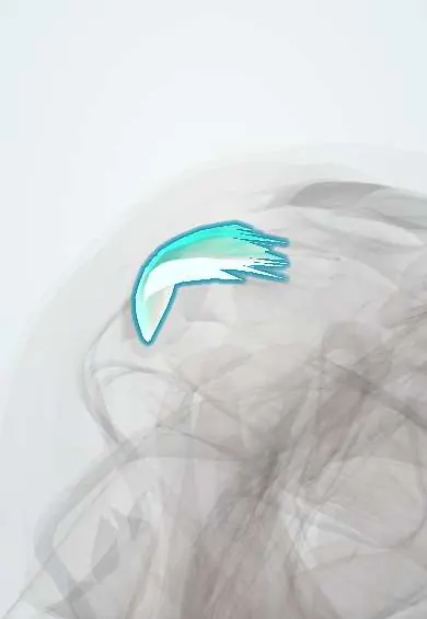
Supraspinatus tendon !
The supraspinatus tendon runs over the top of the shoulder to the humerus; it helps lift the arm and is the rotator cuff tendon most often torn.
Opens in the Durgical app, no sign-up needed.
What it is
The rotator cuff tendon on top of the shoulder, running under the acromion to the greater tuberosity.
What it does
Helps lift the arm out to the side and keeps the ball centred in the socket.
Key facts
- Attaches
- Supraspinatus to the superior facet of the greater tubercle, passing under the acromion and coracoacromial arch; blends with the capsule
- Function
- Starts abduction and holds the humeral head down in the glenoid; the most often torn rotator cuff tendon, usually close to its insertion
- Tight in
- Adduction with medial rotation (hand behind the back)
Relations
- Course
- From the supraspinous fossa it runs laterally under the acromioclavicular joint, the acromion and the coracoacromial ligament, over the top of the humeral head, to the greater tubercle
- Superficial
- Subacromial bursa, then the acromion, the coracoacromial ligament and the deltoid
- Deep
- Shoulder capsule, with which it blends, and the top of the humeral head
- In front
- Rotator interval, holding the long biceps tendon and the coracohumeral ligament, which separates it from the subscapularis tendon
- Behind
- Infraspinatus tendon, whose fibres interlace with it near the greater tubercle
How to find it
With the hand placed behind the back (shoulder extended and medially rotated), the insertion rolls forward from under the acromion and is felt just in front of and below the anterolateral corner of the acromion; tenderness there points to the tendon. The same position is used to scan it with ultrasound.
Examination
- Jobe (empty can) test: arm raised to about 90° in the plane of the scapula, thumb down, pushed down against resistance; pain suggests tendinopathy, weakness suggests a tear
- Full can test: the same with the thumb up, often less painful; weakness also suggests a tear
- Drop arm test: the arm, raised passively to the side, cannot be lowered slowly and falls; suggests a large full-thickness cuff tear
- Painful arc between about 60° and 120° of abduction, as the tendon passes under the acromion; suggests impingement or tendinopathy
- Neer and Hawkins–Kennedy signs: pain when the tendon is pressed under the arch by forced forward flexion, or flexion with medial rotation; suggest impingement, not a tear itself
On imaging
Ultrasound and MRI both show partial and full-thickness tears. On fat-suppressed coronal MRI a full-thickness tear is fluid crossing the tendon; the coronal images show how far the stump has retracted and the sagittal images show muscle wasting and fat. X-rays show calcific deposits, acromial spurs and, in massive tears, a high-riding humeral head.
Common problems
- Rotator cuff tear, partial or full, from wear with age or from a fall; the most common cause of shoulder weakness.
- Tendinopathy and impingement: pain lifting the arm and at night.
- Calcific tendinitis: calcium deposits that cause sudden severe pain.
How it is diagnosed
Weakness and pain on cuff tests (for example the empty-can test). Ultrasound or MRI shows the tear size and muscle quality.
Treatment
- Physical therapy, pain medicines and sometimes injections, especially for partial and age-related tears.
- Arthroscopic rotator cuff repair with suture anchors for many full-thickness tears, especially recent ones in active people.
- Reverse shoulder replacement for large, irreparable tears with arthritis.
Clinical notes
- Massive tears with a retracted stump and fatty muscle may be irreparable and can progress to cuff tear arthropathy, with the humeral head migrating up against the acromion
- A weak cuff with an intact tendon on imaging raises suprascapular nerve entrapment, at the suprascapular notch or the spinoglenoid notch
- A hooked or curved acromion (Bigliani types II–III) is linked to impingement and tears; a subacromial decompression removes the spur
- Anterior shoulder dislocation in older adults often tears this tendon; weakness after reduction calls for a cuff assessment
Anatomical variants
- An aponeurotic expansion of the tendon may reach toward the intertubercular groove; one MRI series found it in about half of shoulders
Development
Its footprint is on the greater tubercle, which ossifies from its own centre in early childhood and merges with the humeral head long before the proximal physis closes in late adolescence; repeated throwing while that physis is open can widen it (Little Leaguer's shoulder).
Memory aid
SITS for the cuff: Supraspinatus, Infraspinatus and Teres minor on the greater tubercle from top to back, Subscapularis alone on the lesser tubercle.
Test yourself
Think of the answer, then tap the question to see it.
Where does the supraspinatus tendon insert, and what does it do?
On the superior facet of the greater tubercle; it starts abduction and holds the humeral head down in the glenoid.
Where do supraspinatus tendon tears usually begin?
At the front of its insertion, next to the biceps interval, in a poorly supplied critical zone just proximal to the bone.
What does the Jobe (empty can) test show about the supraspinatus tendon?
Arm at 90° in the scapular plane, thumb down, pushed down: pain suggests tendinopathy, weakness suggests a tear.
Which partial-thickness supraspinatus tendon tears are the most common?
Articular-side tears (versus bursal-side or within the tendon).
What is the painful arc of the supraspinatus tendon?
Pain between about 60° and 120° of abduction as the tendon passes under the acromion; suggests impingement or tendinopathy.
Educational information, not medical advice. For any symptom, see a health professional.
Its names
- Official Latin (TA2)
- tendo musculi supraspinati TA2 2457
- Official Spanish (DeCS)
- Tendón del músculo supraespinoso
- English (TA2)
- Supraspinatus tendon
Related
Sources: Terminologia Anatomica (TA2); Anatomy, Shoulder and Upper Limb, Arm Supraspinatus Muscle; Anatomy, Rotator Cuff.
Spotted a mistake?We are always improving for the community. Tell us what is wrong and our team will review it.Report a mistake
Representative model, for educational purposes only.