Cardiovascular system · Lower limb
Top-of-foot arteries !
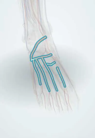
The top-of-foot arteries are the dorsalis pedis artery and its branches; its pulse can be felt on top of the foot.
Official name: Arcuate artery
Opens in the Durgical app, no sign-up needed.
What it is
The dorsalis pedis artery, the continuation of the anterior tibial artery, and its branches on the top of the foot.
What it does
Supply the top of the foot and the toes. The pulse on top of the foot is checked to assess blood flow.
Key facts
- Origin
- Dorsalis pedis, continuation of anterior tibial artery
- Branches
- Lateral and medial tarsal, arcuate (gives dorsal metatarsals 2–4), 1st dorsal metatarsal, deep plantar
- Supplies
- Dorsum of foot and toes; joins plantar arch through deep plantar artery
Relations
- Course
- Lateral tarsal: leaves the dorsalis pedis artery over the navicular and runs laterally under the extensor digitorum brevis toward the cuboid; arcuate: arises near the first tarsometatarsal joint and runs laterally across the bases of metatarsals 2–4, deep to the extensor tendons
- Superficial
- The extensor digitorum longus and extensor digitorum brevis tendons, branches of the superficial fibular nerve and the dorsal venous network
- Deep
- The tarsal bones, the metatarsal bases and the dorsal interossei
How to find it
The dorsalis pedis pulse is the starting point. From it the arcuate artery runs laterally across the metatarsal bases under the extensor tendons, and the first dorsal metatarsal artery runs forward in the first intermetatarsal space; a handheld Doppler follows both.
On imaging
Doppler ultrasound and CT angiography show the arcuate artery and the dorsal metatarsal arteries arising from the dorsalis pedis. Before a toe-to-hand transfer, CT or catheter angiography maps the first dorsal metatarsal artery and its depth relative to the first dorsal interosseous. In peripheral arterial disease, angiography shows how far flow reaches into the foot.
Common problems
- A weak or missing pulse can be a sign of peripheral artery disease.
Clinical notes
- In diabetic foot disease, an intact pedal arch improves the results of distal bypass and wound healing
- The dorsum of the foot is the territory (angiosome) of the anterior tibial artery; some surgeons aim revascularization at the artery that feeds the wound's territory
Anatomical variants
- The arcuate artery is small or absent in many feet (study figures vary widely); the dorsal metatarsal arteries then come from the lateral tarsal artery or from perforating branches of the plantar arch
- The branching pattern on the dorsum varies a great deal, including absence of the dorsalis pedis artery itself
Test yourself
Think of the answer, then tap the question to see it.
What are the main branches of the dorsalis pedis artery on the dorsum of the foot?
Lateral and medial tarsal, arcuate (giving dorsal metatarsals 2–4), first dorsal metatarsal and deep plantar arteries.
How are the dorsal and plantar arterial systems of the foot connected?
Through the deep plantar artery and perforating branches in the intermetatarsal spaces, giving the forefoot a double supply.
Which top-of-foot artery is the key vessel in toe-to-hand transfer?
The first dorsal metatarsal artery; its depth relative to the first dorsal interosseous varies and is mapped before surgery.
Educational information, not medical advice. For any symptom, see a health professional.
Its names
- Official Latin (TA2)
- arteria arcuata; arteria tarsea lateralis TA2 4717
- Official Spanish (DeCS)
- Arterias arcuata y tarsiana lateral
- English (TA2)
- Arcuate artery
Related
- Deep fibular nerve
- Toe arteries
- Extensor digitorum brevis
- Extensor hallucis brevis
- Dorsal interossei
- Talus
- Navicular
Sources: Terminologia Anatomica (TA2); Anatomy, Bony Pelvis and Lower Limb: Foot Arteries.
Spotted a mistake?We are always improving for the community. Tell us what is wrong and our team will review it.Report a mistake
Representative model, for educational purposes only.