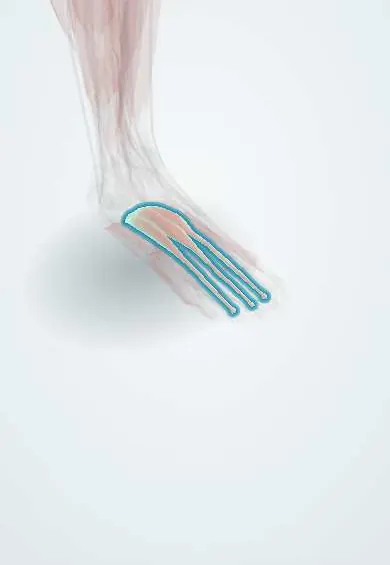
Extensor digitorum brevis !
The extensor digitorum brevis is a small muscle on top of the foot that helps lift the toes; its soft bulge is normal.
Opens in the Durgical app, no sign-up needed.
What it is
A small, flat muscle on top of the foot, starting on the heel bone. With the extensor hallucis brevis, it is the only muscle on top of the foot.
What it does
Helps the long tendons lift the toes.
Key facts
- Origin
- Superolateral calcaneus (floor of sinus tarsi); inferior extensor retinaculum
- Insertion
- Long extensor tendons of toes 2–4
- Action
- Helps extend toes 2–4
- Nerve
- Deep fibular nerve (L5–S1) (some texts: S1–S2)
- Blood supply
- Dorsalis pedis artery (lateral tarsal branch); perforating branch of fibular artery
Relations
- Deep
- Calcaneus and the sinus tarsi, the lateral tarsal bones and the lateral tarsal artery
- Superficial
- Extensor digitorum longus and fibularis tertius tendons, the inferior extensor retinaculum and dorsal branches of the superficial fibular nerve
- Medial
- Extensor hallucis brevis (its medial part), then the dorsalis pedis artery and the deep fibular nerve
- Lateral
- The fibular tendons, which curve below the lateral malleolus behind and below the muscle
How to find it
With the toes extended, a soft, rounded belly appears on the dorsolateral foot just in front of and below the lateral malleolus, over the sinus tarsi.
Examination
- Nerve conduction: a small or absent motor response recorded over it suggests a common or deep fibular nerve lesion; extra response on stimulating behind the lateral malleolus points to an accessory deep fibular nerve
- Wasting, seen as a hollow in front of the lateral malleolus, suggests a long-standing fibular nerve or L5–S1 root problem
On imaging
Ultrasound and MRI show it as a small muscle belly on the dorsolateral foot, in front of the lateral malleolus over the sinus tarsi. Its normal bulge is often referred for a suspected soft-tissue mass, and imaging confirms ordinary muscle. Fatty atrophy of the belly on MRI supports a chronic deep fibular nerve lesion.
Clinical notes
- It is lifted from its origin in the lateral (sinus tarsi) approach to the subtalar joint and calcaneus
- After resection of a calcaneonavicular coalition, it is often placed into the gap to stop the bar from re-forming
- Can be used as a small local muscle flap for defects over the lateral ankle
Anatomical variants
- An accessory deep fibular nerve, a branch of the superficial fibular nerve passing behind the lateral malleolus, supplies part of the muscle in some people
Test yourself
Think of the answer, then tap the question to see it.
What are the origin and nerve supply of the extensor digitorum brevis?
Superolateral calcaneus (floor of the sinus tarsi) and inferior extensor retinaculum; deep fibular nerve.
Why is the extensor digitorum brevis important in nerve conduction studies?
It is the usual recording site for motor conduction of the fibular nerve; a small or absent response suggests a common or deep fibular lesion.
What variant nerve may also supply the extensor digitorum brevis?
An accessory deep fibular nerve, a branch of the superficial fibular nerve passing behind the lateral malleolus.
Educational information, not medical advice. For any symptom, see a health professional.
Its names
- Official Latin (TA2)
- extensor brevis digitorum TA2 2671
- Official Spanish (DeCS)
- Músculo extensor corto de los dedos
- English (TA2)
- Extensor digitorum brevis
- Also called
- extensor digitorum brevis muscle
Related
- Toe extensor tendons
- Extensor hallucis brevis
- Deep fibular nerve
- Top-of-foot arteries
- Calcaneus
- Cuboid
- Lateral cuneiform
- Second metatarsal
Sources: Terminologia Anatomica (TA2); Anatomy, Bony Pelvis and Lower Limb, Foot Muscles.
Spotted a mistake?We are always improving for the community. Tell us what is wrong and our team will review it.Report a mistake
Representative model, for educational purposes only.