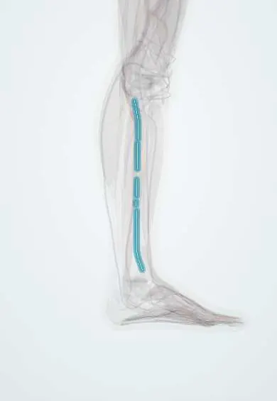
Superficial fibular nerve !
The superficial fibular nerve supplies the outer leg muscles that turn the foot outward and the skin on top of the foot.
Opens in the Durgical app, no sign-up needed.
What it is
A branch of the common peroneal nerve. It controls the outer leg muscles, then comes through the fascia above the ankle to the top of the foot.
What it does
Turns the foot outward (through the fibularis muscles) and carries feeling from most of the top of the foot.
Key facts
- Roots
- L4–S1 (mainly L5–S1)
- Moves
- Lateral leg: fibularis longus and fibularis brevis (eversion)
- Feeling from
- Lower anterolateral leg; most of dorsum of foot except first web space and lateral edge
Relations
- Course
- From the neck of the fibula it descends in the lateral compartment, first between the fibularis longus and the fibula, then between the fibularis longus and brevis, pierces the crural fascia in the distal third of the leg and runs under the skin in front of the ankle to the dorsum of the foot
- In front
- The anterior intermuscular septum and the extensor digitorum longus
- Deep
- The fibula above, then the fibularis brevis
- Superficial
- The fibularis longus above; below its fascial exit, only skin, crossing superficial to the extensor retinaculum
How to find it
With the ankle plantar-flexed and inverted and the fourth toe bent, its intermediate branch often shows as a thin cord under the skin of the anterolateral ankle in slim people. The fascial exit lies on the anterolateral leg some way above the lateral malleolus.
Examination
- Resisted eversion with the foot plantar-flexed: weakness points to this nerve or the L5–S1 roots
- Pinprick over the dorsum of the foot, away from the first web space and the lateral border
- Pressure over the fascial exit while the person dorsiflexes and everts against resistance reproduces pain or tingling in fascial entrapment
On imaging
A thin nerve best seen on high-resolution ultrasound, followed down the lateral compartment between the fibularis muscles and the extensor digitorum longus to where it pierces the deep fascia above the ankle. Ultrasound shows entrapment, a muscle hernia at that exit point or a neuroma after surgery, and guides blocks. On MRI, denervation oedema limited to fibularis longus and brevis points to it.
Common problems
- It can be pinched where it passes through the fascia, or irritated by ankle sprains or ankle surgery.
Clinical notes
- Entrapment at the fascial exit after inversion sprains, in runners, or with a fascial defect and muscle hernia
- Mapping its branches on the skin before placing the anterolateral ankle arthroscopy portal lowers the risk of cutting them
- Blocked by a line of subcutaneous infiltration across the front of the ankle in an ankle block
Anatomical variants
- Most often it pierces the crural fascia and then divides (about 4 in 5 limbs); in about 1 in 6 it divides first and two branches emerge separately; rarely only the medial dorsal cutaneous branch emerges
- Part of its course may run in the anterior instead of the lateral compartment, which matters in fasciotomy
Development
The fibularis longus and brevis it supplies belong, like the anterior compartment, to the dorsal (extensor) mass of the leg bud, so both compartments are served by branches of the common fibular nerve from the posterior divisions of the plexus.
Related conditions
Test yourself
Think of the answer, then tap the question to see it.
What does an isolated superficial fibular nerve lesion cause?
Weak eversion (fibularis longus and brevis) without foot drop, with numbness over most of the dorsum of the foot.
Where is the superficial fibular nerve typically entrapped?
Where it pierces the deep fascia in the lower third of the anterolateral leg, for example after inversion sprains or with a muscle hernia.
Which ankle arthroscopy portal puts the superficial fibular nerve at risk?
The anterolateral portal, because its branches cross the front of the ankle just under the skin; mapping them on the skin lowers the risk.
Educational information, not medical advice. For any symptom, see a health professional.
Its names
- Official Latin (TA2)
- nervus fibularis superficialis TA2 6574
- Official Spanish (DeCS)
- Nervio fibular superficial DeCS D010543
- English (TA2)
- Superficial fibular nerve
- Also called
- superficial peroneal nerve · nervus peronaeus superficialis
Related
Sources: Terminologia Anatomica (TA2); DeCS; Anatomy, Bony Pelvis and Lower Limb: Superficial Peroneal Nerve (Superficial Fibular Nerve).
Spotted a mistake?We are always improving for the community. Tell us what is wrong and our team will review it.Report a mistake
Representative model, for educational purposes only.