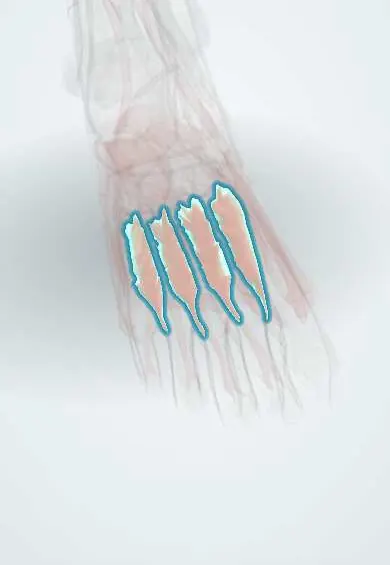
Dorsal interossei !
The dorsal interossei of the foot are four muscles between the metatarsals that spread the toes apart.
Official name: Dorsal interossei muscles of foot
Opens in the Durgical app, no sign-up needed.
What it is
Four muscles in the spaces between the metatarsals.
What it does
Spread the toes apart (away from the second toe) and help bend the toes at the base.
Key facts
- Origin
- Four bipennate muscles from adjacent sides of metatarsals 1–5
- Insertion
- Proximal phalanges and extensor expansions: first to medial side of toe 2; second to fourth to lateral sides of toes 2–4
- Action
- Abduct toes 2–4 from axis through second toe (DAB); flex MTP joints
- Nerve
- Lateral plantar nerve (S2–S3) (varies: first two also from deep fibular nerve)
- Blood supply
- Dorsal metatarsal arteries (arcuate artery); plantar arch perforating branches
Relations
- Above
- Dorsal fascia, the long and short extensor tendons and the dorsal metatarsal arteries
- Below
- Plantar interossei, adductor hallucis, the deep plantar arch and the deep branch of the lateral plantar nerve
- Contents
- Perforating arteries between the two heads of each muscle; in the first space, the deep plantar branch of the dorsalis pedis artery
- Course
- Their tendons run forward, dorsal to the deep transverse metatarsal ligament, to the proximal phalanges and extensor expansions
How to find it
The first dorsal interosseous is felt as a soft fullness in the first intermetatarsal space on the dorsum; the others lie between the metatarsal shafts and are felt only as tissue between the bones.
On imaging
MRI shows them filling the intermetatarsal spaces, best on short-axis images across the metatarsals. Look for oedema after trauma or overuse and for masses between the metatarsal heads, where ultrasound or MRI tells a Morton neuroma from an intermetatarsal bursitis. Radiographs show only the bones; a widened gap between the first and second metatarsal bases suggests a Lisfranc injury.
Common problems
- Like the other small muscles of the foot, it can weaken with age, nerve problems or tight shoes, which can let the arch drop or the toes change shape.
Clinical notes
- Foot compartment syndrome after crush injuries and Lisfranc or calcaneal fractures involves the interosseous compartments; dorsal incisions over the second and fourth metatarsals reach them
- In Lisfranc injuries the deep plantar artery between the first and second metatarsals can be torn, with heavy bleeding
- Their weakness contributes to claw toes
Development
Although they sit between the metatarsals and can be felt from the dorsum, they come from the ventral (flexor) muscle mass of the limb bud, which is why their main nerve supply is the lateral plantar nerve, a branch of the tibial nerve.
Memory aid
"DAB, PAD": Dorsal ABduct, Plantar ADduct, with the second toe as the axis.
Test yourself
Think of the answer, then tap the question to see it.
From which axis do the dorsal interossei of the foot abduct the toes?
The axis through the second toe, not the third as in the hand («DAB»: dorsal abduct).
Why can the second toe move to either side?
It has two dorsal interossei and no plantar interosseous.
Which artery passes between the two heads of the first dorsal interosseous?
The deep plantar branch of the dorsalis pedis artery, which joins the plantar arch.
Educational information, not medical advice. For any symptom, see a health professional.
Its names
- Official Latin (TA2)
- musculi interossei dorsales pedis TA2 2686
- Official Spanish (DeCS)
- Músculos interóseos dorsales del pie
- English (TA2)
- Dorsal interossei muscles of foot
- Also called
- dorsal interossei muscles 1-4 of foot
Related
- Plantar interossei
- Lateral plantar nerve
- Top-of-foot arteries
- Plantar arteries
- First metatarsal
- Second metatarsal
- Third metatarsal
- Fourth metatarsal
Sources: Terminologia Anatomica (TA2); Anatomy, Bony Pelvis and Lower Limb, Foot Muscles.
Spotted a mistake?We are always improving for the community. Tell us what is wrong and our team will review it.Report a mistake
Representative model, for educational purposes only.