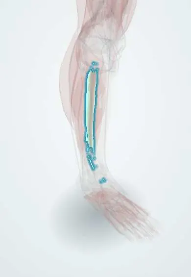
Extensor digitorum longus !
The extensor digitorum longus is a muscle on the front outer leg that lifts the four smaller toes and helps lift the foot.
Opens in the Durgical app, no sign-up needed.
What it is
A muscle on the front of the leg.
What it does
Lifts the toes and the foot.
Key facts
- Origin
- Lateral tibial condyle; upper three-quarters of medial surface of fibula; interosseous membrane
- Insertion
- Middle and distal phalanges of lateral four toes via extensor expansions
- Action
- Extends lateral four toes; dorsiflexes ankle
- Nerve
- Deep fibular nerve (L5–S1)
- Blood supply
- Anterior tibial artery
Relations
- Medial
- Tibialis anterior above and extensor hallucis longus below, with the anterior tibial vessels and deep fibular nerve between them on the interosseous membrane
- Lateral
- Fibularis longus and brevis, beyond the anterior intermuscular septum
- Deep
- Fibula and interosseous membrane
- Superficial
- Deep fascia; in the lower leg the superficial fibular nerve pierces the fascia and runs down over its tendons to the dorsum of the foot
- Course
- Its tendon shares a sheath with fibularis tertius under the extensor retinacula, then splits into four tendons that fan out to toes 2–5, joined by the short extensor tendons for toes 2–4
How to find it
With the toes lifted against light resistance, its four tendons fan out across the dorsum of the foot lateral to the big-toe tendon; at the ankle they lie lateral to the extensor hallucis longus tendon.
Examination
- Resisted extension of the lesser toes: weakness suggests a deep fibular nerve or L5 root problem
- In anterior compartment syndrome, passively curling the toes down stretches the muscle and causes marked pain
On imaging
On axial MRI it is the most lateral muscle of the anterior compartment, against the fibula and the interosseous membrane. Denervation oedema in it and in tibialis anterior, with normal fibularis muscles, points to a deep fibular nerve lesion; adding the fibularis muscles points to the common fibular nerve. Its tendons on the foot are checked with ultrasound for tears or tenosynovitis.
Clinical notes
- Anterior compartment syndrome after tibial fractures or crush injuries involves it together with tibialis anterior and extensor hallucis longus
- Fibular neck fractures and lateral knee approaches can injure the common fibular nerve and its deep branch close to its origin
- Claw and hammer toes: once the small foot muscles fail, the long extensors pull the MTP joints up while the long flexors bend the interphalangeal joints
Anatomical variants
- Fibularis tertius, its lower lateral part, may be absent or small
Development
From the dorsal (extensor) muscle mass, with the deep fibular nerve; the medial rotation of the limb during the seventh week explains why the toe extensors lie at the front of the leg.
Related conditions
Test yourself
Think of the answer, then tap the question to see it.
What are the actions and nerve supply of the extensor digitorum longus?
It extends the lateral four toes and dorsiflexes the ankle; it is supplied by the deep fibular nerve.
Which muscle is formed by the lowest lateral fibres of the extensor digitorum longus?
The fibularis tertius, which may be absent or small.
How does the extensor digitorum longus contribute to claw and hammer toes?
When the small foot muscles fail, it pulls the MTP joints up while the long flexors bend the interphalangeal joints.
Educational information, not medical advice. For any symptom, see a health professional.
Its names
- Official Latin (TA2)
- extensor longus digitorum TA2 2645
- Official Spanish (DeCS)
- Músculo extensor largo de los dedos
- English (TA2)
- Extensor digitorum longus
- Also called
- extensor digitorum longus muscle
Related
- Fibularis tertius
- Common fibular nerve
- Deep fibular nerve
- Anterior tibial artery
- Toe extensor tendon
- Fibula
- Toe extensor tendons
Sources: Terminologia Anatomica (TA2); Anatomy, Bony Pelvis and Lower Limb: Leg Anterior Compartment; 8.5 Development of the Appendicular Skeleton (OpenStax (Rice University), 2022) · adapted, CC BY 4.0.
Spotted a mistake?We are always improving for the community. Tell us what is wrong and our team will review it.Report a mistake
Representative model, for educational purposes only.