Cardiovascular system · Lower limb
Anterior tibial artery !
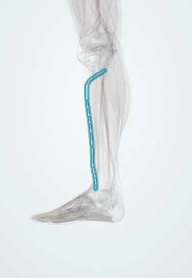
The anterior tibial artery runs down the front of the leg and becomes the dorsalis pedis artery on top of the foot.
Opens in the Durgical app, no sign-up needed.
What it is
An artery that passes to the front of the leg and runs down beside the tibia.
What it does
Supplies the muscles at the front of the leg and continues onto the top of the foot as the dorsalis pedis artery.
Key facts
- Origin
- Popliteal artery at lower border of popliteus; passes above interosseous membrane to front of leg
- Branches
- Anterior and posterior tibial recurrent, muscular, anterior medial and lateral malleolar
- Supplies
- Anterior compartment of leg; continues as dorsalis pedis in front of ankle
Relations
- Course
- From the popliteal bifurcation at the lower border of the popliteus it passes forward between the heads of the tibialis posterior and above the interosseous membrane, descends on the membrane and then on the front of the tibia, and becomes the dorsalis pedis artery midway between the malleoli
- Medial
- The tibialis anterior; near the ankle, the extensor hallucis longus tendon, which crosses in front of it from lateral to medial
- Lateral
- The extensor digitorum longus and, higher up, the extensor hallucis longus
- Deep
- The interosseous membrane, then the lower tibia and the front of the ankle joint
- Superficial
- Deep fascia and, near the ankle, the extensor retinacula
How to find it
Too deep to feel along most of the leg. Its line runs from a point midway between the tibial tuberosity and the head of the fibula down to the midpoint between the malleoli, where its pulse becomes that of the dorsalis pedis artery.
Examination
- Ankle–brachial index: one of the two ankle pressures is taken over the dorsalis pedis, its continuation; after a knee dislocation or upper tibial fracture an abnormal value leads to vascular imaging
- A handheld Doppler at the front of the ankle confirms flow when the dorsalis pedis pulse cannot be felt
On imaging
Duplex ultrasound follows it down the interosseous membrane without contrast. CT angiography is the quick study after knee or upper tibial trauma; catheter angiography shows best how it runs on into the dorsalis pedis. Heavy wall calcification can blur both.
Common problems
- Can be injured in fractures of the tibia or knee dislocations.
- Pressure in a tight muscle compartment (compartment syndrome) can cut off its flow.
Clinical notes
- A common target for distal bypass in limb-salvage surgery
- In high tibial osteotomy and surgery on the upper fibula it lies close behind the bone at the interosseous gap
- In diabetes, artery disease often hits the vessels below the knee, this one included, with long blockages; in the angiosome model it feeds the top of the foot
Anatomical variants
- High origin, above the lower border of the popliteus or even at the level of the knee joint
- Hypoplastic or absent, with the perforating branch of the fibular artery supplying the dorsalis pedis artery
- May give rise to the fibular artery (rare)
- A dorsalis pedis pulse that cannot be felt in a minority of healthy people, because the artery is small, absent or off its usual line
Development
The first arterial axis of the leg is the sciatic artery, which continues below the knee as the fibular artery; the femoral route takes over later and the tibial arteries arise from the axis. This explains why the fibular artery can replace a small or absent anterior tibial artery.
Memory aid
Front of the ankle, medial to lateral: «Tom, Harry And Nervous Dick»: Tibialis anterior, extensor Hallucis longus, Artery, Nerve (deep fibular), extensor Digitorum longus.
Related conditions
- Acute compartment syndrome
- Tibial shaft fracture
- Peripheral artery disease
- Acute limb ischaemia
- Popliteal Artery Injury
Test yourself
Think of the answer, then tap the question to see it.
How does the anterior tibial artery reach the front of the leg?
From the popliteal bifurcation at the lower border of the popliteus, it passes forward above the interosseous membrane.
Why is the anterior tibial artery injured in proximal tibial fractures and knee dislocations?
It is tethered where it passes through the gap above the interosseous membrane.
Where does the anterior tibial artery become the dorsalis pedis artery?
In front of the ankle, midway between the malleoli.
Does a palpable foot pulse rule out anterior compartment syndrome?
No; the foot pulses often stay present until late.
Which ankle pressure in the ankle–brachial index depends on the anterior tibial artery?
The dorsalis pedis pressure, taken over its continuation on the top of the foot.
Educational information, not medical advice. For any symptom, see a health professional.
Its names
- Official Latin (TA2)
- arteria tibialis anterior TA2 4708
- Official Spanish (DeCS)
- Arteria tibial anterior DeCS D016909
- English (TA2)
- Anterior tibial artery
Related
- Deep fibular nerve
- Dorsalis pedis artery
- Tibialis anterior
- Extensor digitorum longus
- Extensor hallucis longus
- Fibularis tertius
Sources: Terminologia Anatomica (TA2); DeCS; Anatomy, Bony Pelvis and Lower Limb: Tibial Artery; 20.5 Circulatory Pathways (OpenStax (Rice University), 2022) · adapted, CC BY 4.0; Anatomy of the Human Body (20th ed.): The Anterior Tibial Artery (Lea & Febiger (Gray's Anatomy, 1918), 1918) · adapted, Public domain; Anatomy of the Human Body (20th ed.): Development of the Vascular System (Lea & Febiger (Gray's Anatomy, 1918), 1918) · adapted, Public domain.
Spotted a mistake?We are always improving for the community. Tell us what is wrong and our team will review it.Report a mistake
Representative model, for educational purposes only.