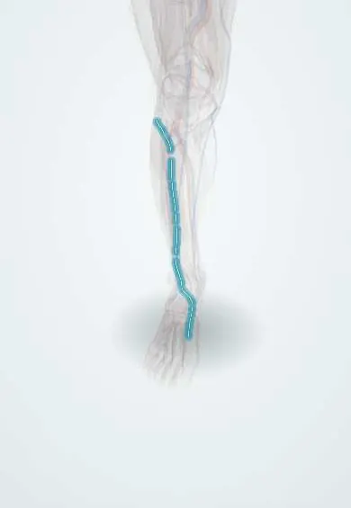
Deep fibular nerve !
The deep fibular nerve supplies the muscles at the front of the leg that lift the foot and toes, and the skin between the first two toes.
Opens in the Durgical app, no sign-up needed.
What it is
A branch of the common peroneal nerve that runs down the front of the leg to the top of the foot.
What it does
Controls the muscles that lift the foot and toes, and carries feeling from the skin between the big toe and the second toe.
Key facts
- Roots
- L4–S1 (mainly L4–L5)
- Moves
- Anterior leg: tibialis anterior, EHL, EDL, fibularis tertius; extensor digitorum brevis and extensor hallucis brevis
- Feeling from
- First web space (adjacent sides of 1st and 2nd toes)
Relations
- Course
- From the neck of the fibula it pierces the anterior intermuscular septum and extensor digitorum longus, descends on the interosseous membrane with the anterior tibial artery, passes under the extensor retinacula and ends on the dorsum as a lateral branch to the extensor digitorum brevis and a medial branch to the first web space
- Medial
- The tibialis anterior in the leg; at the ankle and on the dorsum, the extensor hallucis longus tendon and the dorsalis pedis artery
- Lateral
- The extensor digitorum longus, and in the middle of the leg the extensor hallucis longus before its tendon crosses in front of the nerve
- Deep
- The interosseous membrane, then the front of the distal tibia and the ankle joint capsule
How to find it
It cannot be felt in the leg. At the front of the ankle, lifting the big toe makes the extensor hallucis longus tendon stand out; the nerve runs just lateral to it with the dorsalis pedis artery, whose pulse is the landmark for blocking it.
Examination
- Heel walking and resisted dorsiflexion of the ankle and big toe: weakness with normal eversion points to the deep branch
- Pinprick in the first web space, the one skin area supplied by this nerve alone
- Extensor digitorum brevis bulk on the lateral dorsum: wasting suggests a distal lesion; the muscle is also the usual recording site in nerve conduction studies
On imaging
Too small to judge on radiographs or routine MRI, so it is found by its companion: on ultrasound at the front of the ankle it lies beside the dorsalis pedis artery, between the extensor hallucis longus and extensor digitorum longus tendons. In anterior tarsal tunnel syndrome, radiographs and MRI look for a cause such as dorsal talonavicular osteophytes or a ganglion.
Common problems
- Injury causes foot drop. It can also be pressed by tight laces or straps on top of the foot (anterior tarsal tunnel).
Clinical notes
- Anterior compartment syndrome compresses it early; first-web-space numbness can appear before weakness
- Anterior tarsal tunnel compression also comes from osteophytes or ganglia over the talonavicular joint and from a high boot top
- At risk with anterior ankle approaches, a central anterior arthroscopy portal and pins or screws placed in the distal tibia
- Blocked between the extensor hallucis longus and extensor digitorum longus tendons, next to the dorsalis pedis artery, in an ankle block
Anatomical variants
- Accessory deep fibular nerve: a branch of the superficial fibular nerve that passes behind the lateral malleolus to part of the extensor digitorum brevis; reported in roughly 15–28% of limbs depending on the study, it can keep that muscle working after a deep fibular lesion
Development
Its anterior compartment muscles come from the dorsal (extensor) mass of the leg bud. The lower limb turns medially in week 7 and the upper limb laterally, which is why the extensors sit at the front of the leg but at the back of the forearm.
Related conditions
Test yourself
Think of the answer, then tap the question to see it.
Which skin area does the deep fibular nerve alone supply?
The first web space, between the adjacent sides of the big toe and second toe.
Which muscles does the deep fibular nerve supply?
The anterior compartment (tibialis anterior, extensor hallucis longus, extensor digitorum longus, fibularis tertius) and, on the foot, the extensor digitorum and hallucis brevis.
Where does the deep fibular nerve lie at the front of the ankle?
Between the extensor hallucis longus and extensor digitorum longus tendons, beside the dorsalis pedis artery, whose pulse is the landmark for blocking it.
What is anterior tarsal tunnel syndrome?
Compression of the deep fibular nerve under the inferior extensor retinaculum: first-web-space numbness, sometimes with wasting of the extensor digitorum brevis.
Educational information, not medical advice. For any symptom, see a health professional.
Its names
- Official Latin (TA2)
- nervus fibularis profundus TA2 6579
- Official Spanish (DeCS)
- Nervio fibular profundo DeCS D010543
- English (TA2)
- Deep fibular nerve
- Also called
- deep peroneal nerve · nervus peronaeus profundus
Related
- Common fibular nerve
- Extensor retinacula
- Top-of-foot arteries
- Anterior tibial artery
- Tibialis anterior
- Extensor digitorum longus
- Extensor hallucis longus
- Fibularis tertius
Sources: Terminologia Anatomica (TA2); DeCS; Anatomy, Bony Pelvis and Lower Limb: Calf Deep Peroneal Nerve (Deep Fibular Nerve); 8.5 Development of the Appendicular Skeleton (OpenStax (Rice University), 2022) · adapted, CC BY 4.0.
Spotted a mistake?We are always improving for the community. Tell us what is wrong and our team will review it.Report a mistake
Representative model, for educational purposes only.