Conditions · Ophthalmology and ENT
Otitis media with effusion and chronic suppurative otitis media !
Long-lasting middle ear problems: fluid behind an intact eardrum, or a perforated eardrum that keeps draining. Both cause hearing loss in children.
Updated 2026-11-01
Structures involved
Tap one to see what it is and open it in 3D.
Why it happens
What it is
Two forms of long-standing middle ear disease. Otitis media with effusion (OME) is non-infected fluid behind an intact eardrum without signs of acute infection. Chronic suppurative otitis media (CSOM) is a persistent eardrum perforation with recurrent or continuous ear discharge, usually lasting more than 6 weeks.
Who gets it
OME is very common in young children, often after an episode of acute otitis media or a cold, and most cases clear within weeks to months. CSOM is concentrated in low- and middle-income settings, including rural and Indigenous communities in Latin America, where limited access to care, crowding and malnutrition favour it; it is a leading preventable cause of hearing loss in children worldwide.
Causes
- Eustachian (auditory) tube dysfunction that fails to ventilate and drain the middle ear
- Incompletely resolved or repeated acute otitis media
- Adenoid hypertrophy or a nasopharyngeal mass blocking the tube opening
- CSOM: chronic bacterial infection through a perforation, typically Pseudomonas aeruginosa and Staphylococcus aureus, often mixed and biofilm-forming
Risk factors
- Age under 6, when the auditory tube is short and horizontal
- Day care attendance, bottle feeding while lying down and exposure to tobacco smoke
- Cleft palate and Down syndrome (abnormal tube function)
- Allergic rhinitis and frequent upper respiratory infections
- Poverty, crowding, malnutrition and poor access to care (CSOM)
- In an adult, a new one-sided effusion may signal a nasopharyngeal tumour
Pathophysiology
When the auditory tube does not open well, gas in the middle ear is absorbed, pressure becomes negative and a transudate, later thick mucoid fluid, fills the cavity. The fluid damps eardrum and ossicle movement and causes conductive hearing loss. In CSOM, a perforation lets bacteria from the ear canal colonise the mucosa, which stays inflamed and keeps producing discharge; long-standing disease erodes the ossicles and can lead to cholesteatoma.
Classification
- Otitis media with effusion (serous or mucoid, glue ear): intact eardrum, no acute inflammation
- Chronic suppurative otitis media, mucosal type: central perforation with discharge
- Chronic otitis media with cholesteatoma (squamous type): marginal or attic perforation, see cholesteatoma
- Inactive chronic otitis media: dry perforation
Symptoms and signs
- OME: muffled hearing, ear fullness, often no pain; in small children, speech delay, inattention or poor school performance
- OME on otoscopy: dull, retracted or amber eardrum, sometimes with air-fluid levels or bubbles, and reduced mobility
- CSOM: painless, intermittent or continuous ear discharge (otorrhoea) through a perforation
- CSOM: conductive hearing loss, worse with larger perforations or ossicle damage
Diagnosis
- Clinical
- Pneumatic otoscopy is the key examination: reduced eardrum mobility confirms effusion; in CSOM the size and site of the perforation and the type of discharge are noted. Tuning-fork tests (Rinne, Weber) suggest conductive loss.
- Tests
- Tympanometry shows a flat (type B) curve with effusion. Pure-tone audiometry quantifies hearing loss and is indicated when effusion persists about 3 months. Culture of the discharge guides treatment in resistant CSOM.
- Imaging
- Not routine. CT of the temporal bone when cholesteatoma or a complication is suspected; nasopharyngoscopy, and MRI if needed, for unilateral effusion in an adult.
Differential diagnosis
- Acute otitis media (bulging eardrum, pain and fever)
- Cholesteatoma
- Otitis externa (discharge from the canal skin, pain on pulling the auricle)
- Nasopharyngeal carcinoma in an adult with unilateral effusion
- Sensorineural hearing loss
Goal of treatment
Restore hearing and middle ear ventilation, protect speech and language development in children, dry the ear in CSOM, and detect cholesteatoma and complications early.
Treatment
- Follow-up
- OME: watchful waiting with repeat examination and tympanometry for about 3 months, since most effusions resolve; hearing testing when it persists. Antibiotics, decongestants, antihistamines and nasal steroids are not effective for OME.
- Surgical
- Tympanostomy (ventilation) tubes for persistent bilateral effusion with hearing loss or for children at risk of speech or learning problems; adenoidectomy is added in older children or when tubes have to be repeated.
- Medical
- CSOM: careful cleaning of the ear canal (aural toilet) plus topical antibiotic drops, usually a quinolone, which avoid the ototoxicity of aminoglycoside drops through an open eardrum; systemic antibiotics are reserved for complicated cases.
- Surgical
- Tympanoplasty to close a perforation once the ear is dry, with ossicular reconstruction when the chain is damaged; mastoidectomy when disease extends to the mastoid.
- Follow-up
- Nursing care after tubes: keep water out of the ear when advised, report discharge, watch for tube extrusion, and arrange follow-up hearing tests.
Complications
- Conductive hearing loss and delayed speech and language
- Tympanosclerosis, eardrum retraction pockets and atelectasis
- Ossicular erosion, most often of the incus
- Acquired cholesteatoma
- Mastoiditis, facial nerve palsy and labyrinthitis
- Intracranial spread: meningitis, brain abscess and sigmoid sinus thrombosis
Prognosis
OME usually resolves on its own, and tubes give rapid hearing improvement when it does not. CSOM can be controlled with cleaning and topical drops and closed surgically, but untreated disease leads to permanent hearing loss and occasional serious complications.
Prevention
- Avoiding exposure to tobacco smoke and feeding babies upright
- Breastfeeding and routine childhood immunisation, including pneumococcal vaccine
- Timely treatment and follow-up of acute otitis media
- Hearing screening in children with recurrent ear disease
Test yourself
Think of the answer, then tap the question to see it.
Which tympanometry curve is typical of otitis media with effusion?
A flat (type B) curve, reflecting fluid behind an immobile eardrum.
Which two bacteria most often cause chronic suppurative otitis media?
Pseudomonas aeruginosa and Staphylococcus aureus.
Which ear drops are preferred through a perforated eardrum in CSOM?
Topical quinolone drops, because aminoglycoside drops can be ototoxic.
An adult has new unilateral serous otitis media. Which cancer should be excluded?
Nasopharyngeal carcinoma, by nasopharyngoscopy.
Related conditions
- Acute otitis media
- Cholesteatoma
- Acute mastoiditis
- Hearing loss (conductive and sensorineural, including sudden hearing loss)
- Nasopharyngeal carcinoma
- Tonsillar hypertrophy and recurrent tonsillitis (post-tonsillectomy care)
- Cleft lip and palate
- Down syndrome (trisomy 21)
- Otitis externa
Educational information, not medical advice. For any symptom, see a health professional.
Sources: Otitis media with effusion (MedlinePlus, US National Library of Medicine, 2026); Ear infection - chronic (MedlinePlus, US National Library of Medicine, 2026).
Spotted a mistake?We are always improving for the community. Tell us what is wrong and our team will review it.Report a mistake
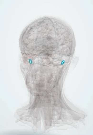
Representative model, for educational purposes only.