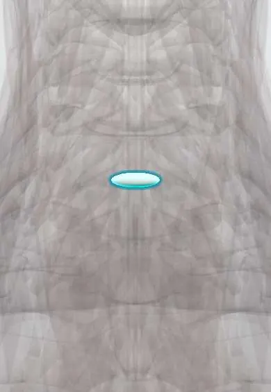
C6–C7 nucleus !
The nucleus pulposus is the soft, gel-like centre of the C6-C7 disc in the neck; it works like a water cushion that spreads the load.
Official name: C6-C7
Opens in the Durgical app, no sign-up needed.
What it is
The soft, gel-like centre (nucleus pulposus) of the C6–C7 disc.
What it does
Works like a water cushion: it turns pressure from above into outward push on the fibre ring, spreading the load. It is under the most pressure when you bend forward and lift.
Key facts
- Where
- Centre of the C6–C7 disc, slightly toward the back, inside the annulus fibrosus
- Function
- Notochord-derived gel (about 70–90% water, proteoglycans); turns axial load into annulus tension; herniates posterolaterally toward the C7 root
Relations
- Behind
- Annulus fibrosus and posterior longitudinal ligament, then the lower cervical cord
- Lateral
- Uncovertebral joints of C7 and the C7 root; the vertebral artery runs one level higher, and the small transverse foramen of C7 usually carries only veins
How to find it
From the front, just below the cricoid cartilage; from behind, between the C6 spinous process and the vertebra prominens (C7).
Examination
- C7 root: weak elbow extension (triceps) and wrist flexion, numb middle finger, reduced triceps reflex
- Neck distraction test: gently lifting the head relieves the arm pain, which supports a compressed root
On imaging
MRI shows the soft fragment and the C7 root; CT separates bone spur and calcified disc from soft nucleus, which matters when planning surgery.
Common problems
- Squeezes out through a tear in the ring (herniation), usually backward and to one side, toward a nerve root.
- Loses water with age, so the disc gets thinner and stiffer.
How it is diagnosed
On MRI a healthy nucleus is bright on T2 images; a worn one turns dark ('black disc').
Treatment
- In a microdiscectomy only the loose piece that has pushed out is removed; most of the disc stays.
Clinical notes
- Surgery is considered for progressive weakness, signs of cord compression or pain that persists; the fragment is removed through an anterior discectomy
- A soft lateral fragment can also be removed from behind through a small opening at the facet (posterior foraminotomy), which keeps the level mobile and avoids a fusion
Related conditions
Test yourself
Think of the answer, then tap the question to see it.
What is the typical picture of a «soft disc» herniation of the C6–C7 nucleus?
A fragment of nucleus in a younger adult, with sudden arm pain into the middle finger (C7 root).
How does a «hard disc» at C6–C7 differ from a soft herniation of the nucleus, and which test tells them apart?
Hard disc: bone spurs and a calcified annulus in older adults, with a slower onset. CT separates spur and calcified disc from soft nucleus.
What is the usual course of an acute C7 radiculopathy from a soft C6–C7 nucleus herniation?
Most improve without surgery over weeks to months.
When is surgery considered for a C6–C7 nucleus herniation, and how is the fragment removed?
For progressive weakness, signs of cord compression or persistent pain; the fragment is removed through an anterior discectomy.
Educational information, not medical advice. For any symptom, see a health professional.
Its names
- Official Latin (TA2)
- nucleus pulposus C6-C7 TA2 1686
- Official Spanish (DeCS)
- Núcleo pulposo C6-C7 DeCS D000070614
- English (TA2)
- C6-C7
Related
Sources: Terminologia Anatomica (TA2); DeCS; Anatomy, Back, Nucleus Pulposus; Anatomy, Back, Intervertebral Discs.
Spotted a mistake?We are always improving for the community. Tell us what is wrong and our team will review it.Report a mistake
Representative model, for educational purposes only.