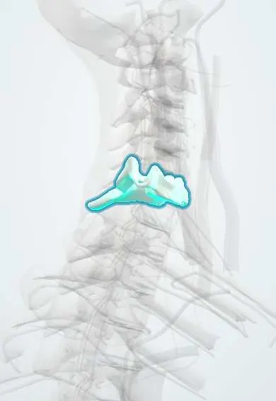
C6 vertebra !
C6 is the sixth neck (cervical) vertebra; with the others it holds up the head, protects the spinal cord and lets the neck move.
Official name: C6
Opens in the Durgical app, no sign-up needed.
What it is
The C6 neck vertebra: a small body in front, a ring behind for the spinal cord, and holes on each side for the vertebral artery.
What it does
Stacks with the other neck vertebrae to hold up the head, protect the spinal cord and let the neck bend and turn.
Key facts
- Joints with
- C5 above and C7 below (intervertebral discs, facet joints and uncovertebral joints)
- Landmarks
- Typical cervical; large anterior (carotid) tubercle; vertebral artery usually enters transverse foramen here; level of cricoid cartilage, start of trachea and esophagus
Relations
- In front
- The esophagus and trachea begin here behind the cricoid cartilage; the inferior thyroid artery and the middle cervical ganglion of the sympathetic trunk lie in front of the transverse process at about this level
- Lateral
- Scalenes from the tubercles; the vertebral artery enters the transverse foramen; the C7 nerve root leaves through the C6–C7 foramen
- Behind
- Short, often bifid spine attached to the nuchal ligament, with semispinalis and multifidus over the laminae
- Contents
- Spinal cord at about the C7 segment, in the lower part of the cervical enlargement
How to find it
In front, the cricoid cartilage is felt as a firm ring just below the thyroid cartilage; the carotid tubercle lies at the same level, deep to the sternocleidomastoid, felt by pressing gently back beside the trachea. From behind, the C6 spine is the one that slides forward and fades under the finger when the neck is extended, unlike C7.
On imaging
Lateral view: the prevertebral soft tissue is normally thicker from C6 down, because the esophagus begins here (commonly quoted upper limit about 22 mm in adults at C6). Oblique views or CT show C6–C7 foraminal spurs; radio-opaque swallowed objects show just below the cricopharyngeus.
Common problems
- Wear and bone spurs (cervical spondylosis), most often at C5-C6 and C6-C7.
- Spurs or a disc can narrow the canal (pressing the cord) or a side opening (pinching a nerve root).
- Fractures from falls, car accidents or diving.
How it is diagnosed
X-rays show alignment and spurs. MRI shows discs, nerves and the spinal cord. CT shows bone detail.
Treatment
- Most neck pain settles with time, activity changes, physical therapy and pain medicine.
- Surgery if a nerve or the cord is pressed and symptoms are severe or getting worse, for example ACDF.
Clinical notes
- Stellate ganglion block: the needle is placed on the C6 carotid tubercle and the injection spreads down to the ganglion at C7–T1; a successful block produces Horner syndrome on that side
- Emergency cricothyrotomy goes through the membrane just above the cricoid; a tracheostomy is placed below it, through the upper tracheal rings
- Cricoid pressure pushes the cricoid ring back against C6 to close the esophagus, a classic but debated maneuver during rapid-sequence intubation
Anatomical variants
- The vertebral artery usually enters the transverse foramen of C6; in a minority it enters at C5, C4 or C7 and runs uncovered in front of the lower transverse processes, where it is exposed in anterior surgery and in stellate ganglion blocks
Development
Each vertebra forms from the lower half of one sclerotome joined to the upper half of the next (resegmentation), so the segmental nerves end up running between vertebrae; this is why the neck has eight cervical nerves but seven cervical vertebrae.
Memory aid
C6, the level of 'sixes': cricoid cartilage, start of esophagus and trachea, carotid tubercle, vertebral artery entering, middle cervical ganglion.
Related conditions
Test yourself
Think of the answer, then tap the question to see it.
Which landmark of the C6 vertebra guides a stellate ganglion block?
The carotid (Chassaignac) tubercle; a successful block produces Horner syndrome on that side.
Which tubes begin at the level of the C6 vertebra and the cricoid cartilage?
The esophagus (from the pharynx) and the trachea (from the larynx); the cricopharyngeus here is the first narrowing of the esophagus, where swallowed objects stick.
Which movement is kept after a complete spinal cord injury at C6, and why does it matter?
Wrist extension, which allows a tenodesis grip and a large gain in independence.
At which vertebra does the vertebral artery usually enter the transverse foramen?
The C6 vertebra; in a minority it enters at C7 or a higher level.
Educational information, not medical advice. For any symptom, see a health professional.
Its names
- Official Latin (TA2)
- vertebra cervicalis VI TA2 1032
- Official Spanish (DeCS)
- Vértebra cervical C6 DeCS D002574
- English (TA2)
- C6
Related
- C5–C6 disc
- C6–C7 disc
- Vertebral artery
- Anterior longitudinal ligament
- Endplates
- Neck facet joints
- Interspinales
- Intertransverse ligaments
Sources: Terminologia Anatomica (TA2); DeCS; Anatomy, Head and Neck: Cervical Vertebrae; Anatomy, Back, Cervical Vertebrae.
Spotted a mistake?We are always improving for the community. Tell us what is wrong and our team will review it.Report a mistake
Representative model, for educational purposes only.