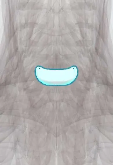
C6–C7 disc !
The C6-C7 disc is the cushion between C6 and C7 in the neck; it is the second most common neck level for a herniated disc.
Official name: C6-C7
Opens in the Durgical app, no sign-up needed.
What it is
The cushion between C6 and C7, the second most common neck level for herniation (C7 nerve root: pain into the middle finger).
What it does
Absorbs load and lets the neck bend and turn. C5-C6 is the most mobile and most worn neck level.
Key facts
- Where
- Between C6 and C7 bodies; taller in front (cervical lordosis); flanked by uncovertebral joints
- Attaches
- Annulus fibrosus anchored to the endplates and ring apophyses of C6 and C7; anterior and posterior longitudinal ligaments
- Function
- Absorbs and spreads load; posterolateral herniation usually compresses the C7 root (one of the two commonest cervical levels)
Relations
- In front
- Longus colli, esophagus and trachea, with the lower poles of the thyroid gland; the recurrent laryngeal nerve climbs in the tracheoesophageal groove
- Behind
- Posterior longitudinal ligament, the dural sac and the lower cervical spinal cord
- Lateral
- Uncovertebral joint and the C7 nerve root in the C6–C7 foramen; the vertebral artery lies inside the C6 transverse foramen above but in front of the C7 transverse process below
How to find it
From behind, between the C6 spinous process and the vertebra prominens (C7 vertebra). To tell them apart, the C6 spinous process slides forward under the finger when the neck extends, while C7 stays put. From the front it lies just below the cricoid cartilage.
Examination
- Elbow extension against resistance with a reduced triceps reflex: C7 nerve root
- Upper limb tension test (median nerve bias): reproduction of the arm symptoms supports cervical radiculopathy; a negative test makes it less likely
On imaging
A lateral cervical radiograph must show down to the top of T1; if the shoulders hide C6–C7, a swimmer's view or CT is needed. This is one of the most degenerated levels, with disc narrowing and spurs. MRI is the test for radiculopathy: axial T2 shows a disc or spur in the C6–C7 foramen pressing on the C7 root. CT myelography replaces MRI when it is contraindicated.
Common problems
- Herniation pressing on the C6 nerve root: neck pain and pain, numbness or weakness down the arm into the thumb side of the hand.
- Degeneration with bone spurs, which can narrow the canal and press on the spinal cord (myelopathy: clumsy hands, unsteady walking).
How it is diagnosed
Exam of arm strength, reflexes and feeling, plus signs of cord pressure. MRI shows the disc, nerve roots and cord.
Treatment
- Most arm pain from a disc improves with time, physical therapy, medicine and sometimes an injection.
- Lasting arm pain, weakness or cord pressure: anterior cervical discectomy and fusion (ACDF), or disc replacement in selected patients.
Clinical notes
- Carpal tunnel syndrome also numbs the middle finger but spares the triceps and its reflex; nerve conduction studies help separate the two, and both can coexist
- In short or heavy necks the shoulders can hide C6–C7 on the lateral X-ray taken during surgery; gentle traction on the arms brings it into view
Development
The neck curve is a secondary curve: it forms after birth, once the infant starts holding the head up. The lower cervical discs are taller at the front than at the back and help build it, so when C6–C7 dries out and narrows the neck often straightens on the lateral X-ray.
Memory aid
Reflex rhyme: «5, 6, pick up sticks» (biceps), «7, 8, lay them straight» (triceps).
Related conditions
Test yourself
Think of the answer, then tap the question to see it.
What is the clinical picture of the C7 root compressed by a C6–C7 disc herniation?
Weak elbow extension (triceps), wrist flexion and finger extension, a reduced triceps reflex and numbness of the middle finger.
How is C7 radiculopathy from a C6–C7 disc told apart from carpal tunnel syndrome?
Carpal tunnel syndrome also numbs the middle finger but spares the triceps and its reflex; nerve conduction studies help, and both can coexist.
Where does C7 root pain from a C6–C7 disc often spread before reaching the hand?
To the back of the arm and the inner edge of the shoulder blade.
What does the upper limb tension test (median nerve bias) add in suspected C6–C7 radiculopathy?
Reproduction of the arm symptoms supports cervical radiculopathy; a negative test makes it less likely.
Educational information, not medical advice. For any symptom, see a health professional.
Its names
- Official Latin (TA2)
- discus intervertebralis C6-C7 TA2 1684
- Official Spanish (DeCS)
- Disco C6-C7 DeCS D007403
- English (TA2)
- C6-C7
- Also called
- fibrous ring · fibrocartilago intervertebralis
Related
Sources: Terminologia Anatomica (TA2); DeCS; Anatomy, Back, Intervertebral Discs; 7.3 The Vertebral Column (OpenStax (Rice University), 2022) · adapted, CC BY 4.0; Anatomy of the Human Body (20th ed.): Articulations of the Vertebral Column (Lea & Febiger (Gray's Anatomy, 1918), 1918) · adapted, Public domain.
Spotted a mistake?We are always improving for the community. Tell us what is wrong and our team will review it.Report a mistake
Representative model, for educational purposes only.