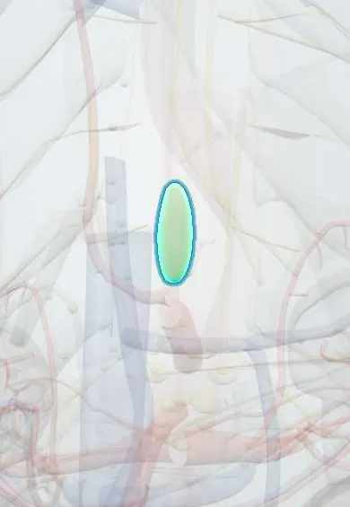
Cisterna chyli !
The cisterna chyli is a small sac at the back of the upper abdomen where lymph from the gut and legs collects before entering the thoracic duct.
Opens in the Durgical app, no sign-up needed.
What it is
A small sac in front of the upper lumbar spine where the gut's lymph vessels meet; the start of the thoracic duct.
What it does
Collects lymph and absorbed fat (chyle) from the intestines.
Key facts
- Where
- Retroperitoneal sac in front of L1–L2, right of aorta, behind right crus of diaphragm (varies: absent or a plexus)
- Drains
- Intestinal trunk (chyle from gut) and right and left lumbar trunks (lower limbs, pelvis, kidneys, abdominal wall)
- Into
- Thoracic duct, which ascends through aortic hiatus (T12)
Relations
- In front
- The right crus of the diaphragm
- Behind
- Bodies of L1 and L2
- Medial
- The abdominal aorta, on its left
- Lateral
- The start of the azygos vein, on its right
- Above
- Continues as the thoracic duct, which passes through the aortic hiatus (T12) between the aorta and the azygos vein
- Below
- Receives the right and left lumbar trunks and the intestinal trunk
How to find it
Not palpable. It lies on the front of the upper lumbar vertebrae, to the right of the aorta and behind the right crus of the diaphragm. For lymphatic embolisation it is first filled with contrast by lymphangiography and then punctured with a fine needle through the abdomen under fluoroscopy.
On imaging
On CT or MRI it may show as a small oval or tubular fluid-filled structure behind the right crus, bright on heavily T2-weighted MRI; it should not be mistaken for a retrocrural lymph node.
Clinical notes
- Chylous ascites: milky, triglyceride-rich peritoneal fluid after retroperitoneal node dissection, aortic or pancreatic surgery; lymphoma and abdominal tuberculosis are other causes
- Virchow node: cancer cells from the abdomen travel up the thoracic duct to the left supraclavicular node (Troisier sign)
- Chyle leaks can be treated by lymphangiography and embolization, reaching the thoracic duct through a needle placed into the cisterna chyli
Development
It forms from one of the primary lymph sacs of the embryo; the thoracic duct develops from channels linking it to the jugular lymph sacs, which explains its frequent variants (doubled duct, plexus instead of a sac).
Test yourself
Think of the answer, then tap the question to see it.
Where is the cisterna chyli?
A retroperitoneal sac in front of L1–L2, right of the aorta and behind the right crus of the diaphragm; in some people it is a plexus instead of a sac.
What drains into the cisterna chyli, and what does it continue as?
The intestinal trunk (chyle from the gut) and the right and left lumbar trunks; it continues as the thoracic duct through the aortic hiatus (T12).
What does injury to the cisterna chyli cause?
Chylous ascites: milky, triglyceride-rich peritoneal fluid, for example after retroperitoneal node dissection or aortic or pancreatic surgery.
Educational information, not medical advice. For any symptom, see a health professional.
Its names
- Official Latin (TA2)
- cisterna chyli TA2 5145
- Official Spanish (DeCS)
- Cisterna del quilo
- English (TA2)
- Cisterna chyli
- Also called
- chyle cistern · cistern of Pecquet
Sources: Terminologia Anatomica (TA2); Anatomy, Abdomen and Pelvis: Lymphatic Drainage; Anatomy of the Human Body (20th ed.): The Abdominal Aorta (Lea & Febiger (Gray's Anatomy, 1918), 1918) · adapted, Public domain.
Spotted a mistake?We are always improving for the community. Tell us what is wrong and our team will review it.Report a mistake
Representative model, for educational purposes only.