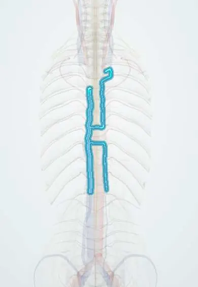
Azygos veins !
The azygos system is a set of veins running up both sides of the thoracic spine; it drains the chest wall and back.
Official name: Azygos vein
Opens in the Durgical app, no sign-up needed.
What it is
Veins running up the right and left sides of the thoracic spine.
What it does
Drain the chest wall and back, linking the two venae cavae.
Key facts
- Drains
- Posterior chest and abdominal wall (posterior intercostal, subcostal, ascending lumbar veins), bronchial and oesophageal veins; hemiazygos veins on the left
- Into
- Azygos arches over the right main bronchus into the superior vena cava at T4; collateral path between IVC and SVC
Relations
- Course
- The azygos vein begins near L1–L2 from the right ascending lumbar and subcostal veins, enters the thorax through the aortic hiatus or the right crus, climbs on the right of the vertebral bodies and arches forward over the root of the right lung at T4 into the superior vena cava
- In front
- Oesophagus and, at the arch, the root of the right lung
- Behind
- Thoracic vertebral bodies and the right posterior intercostal arteries
- Medial
- Thoracic duct and descending thoracic aorta, on its left
- Lateral
- Right mediastinal pleura and right lung
How to find it
It cannot be felt. In a right thoracotomy or thoracoscopy the azygos vein is seen climbing the right side of the vertebral bodies, to the right of the thoracic duct and aorta, then arching forward over the right main bronchus at about T4 to enter the superior vena cava.
On imaging
On a frontal chest radiograph the azygos arch makes a small oval shadow in the right tracheobronchial angle; it enlarges when lying flat, when right heart pressure rises and with azygos continuation of the inferior vena cava.
Clinical notes
- Divided or tied at its arch in right thoracotomy approaches to the oesophagus and the upper thoracic spine
- Large azygos and hemiazygos veins on CT with no hepatic segment of the inferior vena cava mean azygos continuation, which matters before cardiac, liver or retroperitoneal surgery
- Raised abdominal or chest pressure drives blood between the azygos system and the vertebral venous plexuses, one reason epidural veins bleed more when the abdomen is compressed
Anatomical variants
- Azygos lobe of the right lung in about 1 in 200 people (commonly quoted 0.4–1%): the arch runs through the upper lobe inside a pleural fold, seen on the radiograph as a fine curved line
- Azygos continuation of the inferior vena cava: the hepatic segment is missing and the lower body drains through a large azygos vein, often with polysplenia
- The hemiazygos veins vary in size and in how they join the azygos; absence of the azygos vein itself is rare
Development
The azygos vein comes from the right supracardinal vein and the hemiazygos from the left; when the hepatic segment of the inferior vena cava fails to form, the lower body drains through the azygos instead.
Test yourself
Think of the answer, then tap the question to see it.
Where does the azygos vein end?
It arches over the root of the right lung (right main bronchus) at T4 and drains into the superior vena cava.
What is the main collateral role of the azygos system?
It is the main collateral route between the inferior and superior venae cavae when either is blocked.
At what level do the hemiazygos veins cross to join the azygos vein, and behind what?
At about T8–T9, crossing behind the aorta.
What is an azygos lobe?
A variant (about 1 in 200) where the azygos arch runs through the right upper lobe inside a pleural fold, seen as a fine curved line on the radiograph.
Educational information, not medical advice. For any symptom, see a health professional.
Its names
- Official Latin (TA2)
- vena azyga; vena hemiazyga; vena hemiazyga accessoria TA2 4747
- Official Spanish (DeCS)
- Vena ácigos DeCS D001401
- English (TA2)
- Azygos vein
- Also called
- inferior hemiazygos vein · inferior hemi-azygos vein · superior hemiazygos vein · superior hemi-azygos vein · vena thoracica longitudinalis dextra
Related
Sources: Terminologia Anatomica (TA2); DeCS; Anatomy, Thorax, Azygos Veins; 20.5 Circulatory Pathways (OpenStax (Rice University), 2022) · adapted, CC BY 4.0.
Spotted a mistake?We are always improving for the community. Tell us what is wrong and our team will review it.Report a mistake
Representative model, for educational purposes only.