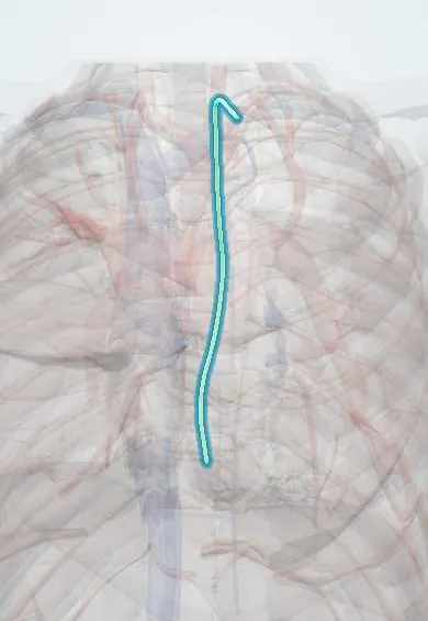
Thoracic duct !
The thoracic duct is the body's main lymph vessel, running up the back of the chest; it returns lymph and absorbed fat to the bloodstream.
Opens in the Durgical app, no sign-up needed.
What it is
The body's largest lymph vessel, running up beside the aorta and esophagus to a vein at the base of the neck.
What it does
Returns lymph and absorbed fat from most of the body to the blood.
Key facts
- Where
- From cisterna chyli (L1–L2) via aortic hiatus (T12); rises between aorta and azygos behind esophagus, crosses left at about T5
- Drains
- Lymph (and absorbed fat) from lower body, left thorax, left upper limb, left head and neck
- Into
- Left venous angle (junction of left subclavian and internal jugular veins)
Relations
- Course
- From the cisterna chyli it passes through the aortic hiatus at T12, ascends on the right between the aorta and azygos vein behind the esophagus, crosses to the left at about T5, climbs on the left of the esophagus into the neck and arches forward to the left venous angle
- In front
- Below T5: the esophagus (and the diaphragm at the hiatus); above T5: the aortic arch and the start of the left subclavian artery
- Behind
- Vertebral bodies, right posterior intercostal arteries, and the hemiazygos and accessory hemiazygos veins crossing to the azygos
- Lateral
- Below T5: the azygos vein on its right and the descending aorta on its left; above T5: the left mediastinal pleura on its left
- Above
- In the root of the neck its arch lies behind the carotid sheath (common carotid artery, internal jugular vein, vagus) and in front of the vertebral vessels, the sympathetic trunk, the thyrocervical trunk and the phrenic nerve on scalenus anterior; it receives the left jugular, subclavian and bronchomediastinal trunks
How to find it
Not palpable. At surgery it is found just above the diaphragm, on the right, behind the esophagus between the descending aorta and the azygos vein; in the neck it is sought at the left venous angle, where the internal jugular and subclavian veins meet. Lymphangiography through groin nodes outlines it for embolization of a leak.
On imaging
Rarely seen on routine CT; the cisterna chyli can appear as a small fluid-filled tube to the right of the aorta at L1–L2. Conventional or MR lymphangiography shows the duct and the site of a leak, and guides percutaneous embolization.
Common problems
- Injury during surgery leaks milky fluid into the chest (chylothorax).
Clinical notes
- Chylothorax after esophagectomy and lung or heart surgery, or from lymphoma; the fluid is milky and rich in triglycerides and chylomicrons
- Ligation just above the diaphragm, between the aorta and the azygos vein, is the classic operation for a leak that persists
- Left neck dissection can injure its termination and cause a chyle leak in the neck
- An enlarged left supraclavicular node near its end (Virchow node) may signal abdominal cancer spreading upward (Troisier sign)
Anatomical variants
- The textbook single-channel course is found in only about half of people; double or plexus-like segments and a right-sided ending occur
- A distinct cisterna chyli is present in only about half of people
Development
Forms from a right and a left embryonic duct joined by cross-links: the lower part of the right duct, a cross-link and the upper part of the left duct usually persist, which explains its switch from right to left and its many double or plexus-like variants.
Related conditions
Test yourself
Think of the answer, then tap the question to see it.
Where does the thoracic duct begin, enter the chest and end?
From the cisterna chyli (L1–L2), through the aortic hiatus (T12), to the left venous angle where the left subclavian and internal jugular veins join.
Why does thoracic duct damage below T5 tend to cause a right chylothorax?
Below about T5 the duct lies on the right, between the aorta and azygos vein; above T5 it crosses to the left, so damage there gives a left chylothorax.
What does chylothorax fluid look like?
Milky, and rich in triglycerides and chylomicrons.
Where is the thoracic duct ligated for a persistent chyle leak?
Just above the diaphragm, between the aorta and the azygos vein.
Educational information, not medical advice. For any symptom, see a health professional.
Its names
- Official Latin (TA2)
- ductus thoracicus TA2 5137
- Official Spanish (DeCS)
- Conducto torácico DeCS D013897
- English (TA2)
- Thoracic duct
Related
Sources: Terminologia Anatomica (TA2); DeCS; Anatomy, Thorax, Thoracic Duct; Tratado de anatomía humana (9.ª ed.), tomo II, Libro IV Angiología, sección 5: Linfáticos.
Spotted a mistake?We are always improving for the community. Tell us what is wrong and our team will review it.Report a mistake
Representative model, for educational purposes only.