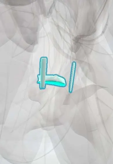
Sacrococcygeal joint !
The sacrococcygeal joint is the small joint between the sacrum and the tailbone (coccyx); it lets the coccyx tilt slightly.
Opens in the Durgical app, no sign-up needed.
What it is
The small joint between the sacrum and the coccyx.
What it does
Lets the tailbone tilt slightly back when you sit and during childbirth.
Key facts
- Where
- Between the apex of the sacrum (S5) and the base of the coccyx (Co1)
- Attaches
- Fibrocartilaginous disc; anterior, posterior (superficial and deep) and lateral sacrococcygeal ligaments; intercornual ligaments
- Function
- Symphysis-type joint; slight flexion-extension, coccyx moves back in sitting and childbirth; often fuses with age
Relations
- In front
- Presacral (retrorectal) space and the rectum; the ganglion impar and the median sacral artery lie on the front of the joint
- Behind
- Superficial and deep posterior sacrococcygeal ligaments; gluteus maximus arises from the side of the lower sacrum and coccyx; the skin of the natal cleft
- Lateral
- Lateral sacrococcygeal ligaments, the coccygeus muscle and the sacrospinous ligament and sacrotuberous ligament along the edges of the lower sacrum and coccyx
- Below
- Coccyx, with the anococcygeal ligament and the levator ani of the pelvic floor attached near its tip
How to find it
In the natal cleft the two sacral cornua are felt as small bumps marking the sacral hiatus; a little lower a slight groove or movable step marks the joint, with the coccyx below. On rectal examination the coccyx can be held between finger and thumb to test movement and tenderness.
Examination
- Pain reproduced by moving the coccyx between a finger in the rectum and a thumb outside, with painless pressure elsewhere, points to coccydynia rather than referred pain
On imaging
MRI shows bone edema, fractures and presacral masses such as chordoma; prenatal ultrasound detects sacrococcygeal teratoma before birth.
Common problems
- Coccydynia: pain at the tailbone when sitting, after a fall, childbirth or for no clear reason.
- Fracture or dislocation from a fall onto the buttocks.
How it is diagnosed
Exam (tenderness at the tailbone); X-rays sitting and standing can show abnormal movement.
Treatment
- A cushion with a cut-out, anti-inflammatory medicine, pelvic floor physical therapy, injections.
- Rarely, removal of the coccyx (coccygectomy) when nothing else helps.
Clinical notes
- Coccygectomy is reserved for persistent coccydynia; wound infection is the main complication because of the nearby anus
- Pilonidal disease in the natal cleft over the coccyx is a skin and hair-follicle problem, not a joint one, but is felt at the same spot
- During vaginal birth the coccyx moves backward at this joint to enlarge the pelvic outlet; a difficult delivery can sprain or dislocate it and is a known cause of coccyx pain afterwards
Anatomical variants
- The joint fuses in many adults, especially with age; in some it is partly synovial
- The coccyx usually has four segments (range three to five); a sharply forward-hooked coccyx is linked to coccydynia
Development
The coccyx is the remnant of the embryonic tail; its segments ossify late, the first in the early years of life and the last around or after puberty. Remnants of the notochord here explain chordoma.
Test yourself
Think of the answer, then tap the question to see it.
Which ganglion lies in front of the sacrococcygeal joint, and why is it targeted?
The ganglion impar (of Walther), where the two sympathetic trunks meet; it is blocked for coccyx and perineal pain.
Which opening just above the sacrococcygeal joint is the entry for a caudal epidural?
The sacral hiatus, between the sacral cornua, closed by the superficial posterior sacrococcygeal ligament.
Which tumor of the sacrococcygeal region is the most common tumor of newborns?
Sacrococcygeal teratoma; prenatal ultrasound can detect it before birth.
Why is coccydynia, felt at the sacrococcygeal joint, more common in women?
It is linked to the wider pelvis, childbirth and a more backward-tilted coccyx; it is several times more common in women.
Educational information, not medical advice. For any symptom, see a health professional.
Its names
- Official Latin (TA2)
- junctura sacrococcygea TA2 1688
- Official Spanish (DeCS)
- Articulación sacrococcígea
- English (TA2)
- Sacrococcygeal joint
- Also called
- articulatio sacrococcygea
Related
Sources: Terminologia Anatomica (TA2); Anatomy, Back, Coccygeal Vertebrae.
Spotted a mistake?We are always improving for the community. Tell us what is wrong and our team will review it.Report a mistake
Representative model, for educational purposes only.