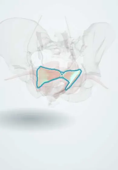
Iliococcygeus !
The iliococcygeus is the thin, flat back part of the levator ani in the pelvic floor; it forms a shelf that supports the pelvic organs.
Official name: Iliococcygeus muscle
Opens in the Durgical app, no sign-up needed.
What it is
The thin back part of the levator ani, from a tendinous arch on the pelvic side wall to the coccyx.
What it does
Forms the flat back of the pelvic floor that supports the organs.
Key facts
- Origin
- Tendinous arch of levator ani (obturator fascia) and ischial spine
- Insertion
- Anococcygeal body and coccyx
- Action
- Forms the posterior pelvic floor plate; supports the viscera
- Nerve
- Nerve to levator ani (S3–S4) and pudendal nerve (S2–S4) branches
- Blood supply
- Inferior gluteal, internal pudendal and inferior vesical/vaginal arteries
Relations
- In front
- Pubococcygeus, with which it often blends without a clear boundary
- Behind
- Coccygeus, which continues the floor back to the sacrum
- Lateral
- Tendinous arch of levator ani on the obturator internus; below the arch the obturator internus forms the outer wall of the ischioanal fossa
- Above
- Pelvic fascia, then the rectum and the pelvic organs that rest on the levator plate
- Below
- Fat of the ischioanal fossa, with the pudendal canal on its lateral wall
How to find it
It cannot be felt from outside. On vaginal or rectal examination it is a thin sheet on the posterolateral wall, higher and less distinct than the puborectalis sling. In surgery it is seen from above when the peritoneum and pelvic fascia are lifted off the side wall.
On imaging
On sagittal and coronal MRI the levator plate normally lies close to horizontal; in pelvic organ prolapse and with ageing it tilts downwards and the floor sags into a deeper bowl, widening the hiatus in front of it.
Common problems
- Stretching or tearing in vaginal birth.
- Pelvic organ prolapse.
- Urinary or faecal incontinence.
- Overactive, painful pelvic floor (pain with sex, pelvic pain).
How it is diagnosed
Pelvic examination, sometimes ultrasound or MRI of the pelvic floor.
Treatment
- Pelvic floor muscle training (Kegel exercises) with a physiotherapist.
- A pessary (a removable support placed in the vagina).
- Surgery for prolapse that bothers the person.
Clinical notes
- Iliococcygeus fascia fixation sutures the vaginal vault to the fascia over this muscle just below and in front of the ischial spine, an alternative to sacrospinous fixation for vault prolapse
- A downward-tilted levator plate is associated with uterine and vault prolapse, because the organs lose the shelf that holds them over the floor
Anatomical variants
- A gap can exist between the muscle and its tendinous arch (hiatus of Schwalbe), a rare site of pelvic floor hernia
- MRI may show it thin or partly absent in some women; with ageing the genital hiatus widens
Related conditions
Test yourself
Think of the answer, then tap the question to see it.
What shelf does the iliococcygeus form with the back fibres of the pubococcygeus, and what rests on it?
The levator plate, a near-horizontal shelf on which the rectum, upper vagina and uterus rest when standing.
From where does the iliococcygeus arise?
From fascia, the back part of the tendinous arch of levator ani, and from the ischial spine; it is the thinnest part of the levator ani.
What is iliococcygeus fascia fixation used for?
Vaginal vault prolapse: the vault is sutured to the fascia over the iliococcygeus just below and in front of the ischial spine.
Educational information, not medical advice. For any symptom, see a health professional.
Its names
- Official Latin (TA2)
- musculus iliococcygeus TA2 2404
- Official Spanish (DeCS)
- Músculo iliococcígeo DeCS D017773
- English (TA2)
- Iliococcygeus muscle
Related
Sources: Terminologia Anatomica (TA2); DeCS; Anatomy, Abdomen and Pelvis: Levator Ani Muscle; Anatomy, Abdomen and Pelvis: Pelvic Floor.
Spotted a mistake?We are always improving for the community. Tell us what is wrong and our team will review it.Report a mistake
Representative model, for educational purposes only.