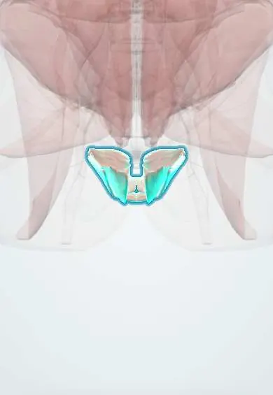
Pelvic floor !
The pelvic floor is a hammock of muscles across the bottom of the pelvis; it supports the pelvic organs and controls the bladder and bowel.
Official name: Levator ani
Opens in the Durgical app, no sign-up needed.
What it is
A hammock of muscles across the bottom of the pelvis, from the pubis to the coccyx.
What it does
Supports the pelvic organs and controls the bladder and bowel; works with the deep abdominal muscles to support the spine.
Key facts
- Origin
- Levator ani: body of pubis, tendinous arch of obturator fascia, ischial spine; coccygeus: ischial spine and sacrospinous ligament
- Insertion
- Perineal body, anococcygeal ligament, coccyx; coccygeus: lower sacrum and coccyx
- Action
- Supports pelvic viscera; resists intra-abdominal pressure; puborectalis keeps the anorectal angle for continence
- Nerve
- Nerve to levator ani (ventral rami S3–S4) and pudendal nerve branches (S2–S4); coccygeus: S3–S4
- Blood supply
- Inferior gluteal, internal pudendal and inferior vesical arteries
Relations
- Above
- Pelvic organs with the endopelvic fascia: bladder, uterus and vagina in women or prostate in men, and rectum
- Below
- Ischioanal fossae on each side, with the pudendal canal in their lateral wall; the perineal membrane and the external genital muscles in front
- Lateral
- Obturator internus and its fascia, from whose tendinous arch levator ani arises
- Behind
- Coccyx and sacrum; piriformis lies just above coccygeus
How to find it
On digital rectal or vaginal examination the puborectalis is felt as a firm sling on the back and sides of the canal a few centimetres in; asking the person to squeeze shows its contraction and inward lift.
Examination
- Digital assessment of pelvic floor contraction, often graded on the modified Oxford scale: a weak or absent squeeze suggests muscle injury or denervation
On imaging
Transperineal 3D ultrasound and dynamic pelvic MRI show avulsion of levator ani from the pubic bone and a hiatus that balloons on straining.
Clinical notes
- Vaginal birth can stretch or tear levator ani (avulsion from the pubic bone, more often after forceps delivery) and stretch the pudendal nerve, raising the later risk of prolapse and incontinence
- Pelvic floor muscle training is the first-line conservative approach to stress urinary incontinence
- Dyssynergic defecation: puborectalis fails to relax or tightens on straining, causing obstructed defecation
- With ageing the hiatus widens and puborectalis thins
Anatomical variants
- Nerve supply varies: some fibres come directly from S4 or from the coccygeal plexus; MRI shows thinning or absence of parts of levator ani in some women
Related conditions
Test yourself
Think of the answer, then tap the question to see it.
What are the three parts of the levator ani in the pelvic floor?
Puborectalis, pubococcygeus and iliococcygeus; the coccygeus lies behind them on the sacrospinous ligament.
What is the role of the puborectalis in continence?
It slings the anorectal junction forward to keep the anorectal angle, and relaxes to allow defecation.
What nerves supply the pelvic floor?
The nerve to levator ani (S3–S4) and branches of the pudendal nerve (S2–S4).
Which obstetric event can injure the pelvic floor, and with what later risk?
Vaginal birth, especially forceps delivery, can avulse levator ani from the pubis and stretch the pudendal nerve, raising later prolapse and incontinence risk.
What is dyssynergic defecation?
The puborectalis fails to relax or tightens on straining, causing obstructed defecation.
Educational information, not medical advice. For any symptom, see a health professional.
Its names
- Official Latin (TA2)
- levator ani; musculus coccygeus TA2 2403
- Official Spanish (DeCS)
- Diafragma pélvico DeCS D017773
- English (TA2)
- Levator ani
- Also called
- levator ani muscle · ischiococcygeus muscle
Related
Sources: Terminologia Anatomica (TA2); DeCS; Anatomy, Abdomen and Pelvis: Pelvic Floor; Anatomy, Abdomen and Pelvis: Levator Ani Muscle.
Spotted a mistake?We are always improving for the community. Tell us what is wrong and our team will review it.Report a mistake
Representative model, for educational purposes only.