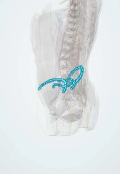
Iliolumbar vessels !
The iliolumbar artery and vein lie near L5 and the inner side of the iliac wing; they supply the psoas, the iliacus and the lower spine.
Official name: Iliolumbar artery
Opens in the Durgical app, no sign-up needed.
What it is
An artery and vein near the L5 transverse process and the inner side of the iliac wing.
What it does
Supply the psoas, iliacus, quadratus lumborum and lower spine. The vein ties the left common iliac vein to the side wall and is tied before moving it in ALIF.
Key facts
- Origin
- Iliolumbar artery: posterior division of internal iliac artery; iliolumbar vein drains into the internal or common iliac vein
- Branches
- Lumbar branch (psoas, quadratus lumborum, spinal branch at L5–S1) and iliac branch (iliacus, ilium)
- Supplies
- Psoas, quadratus lumborum, iliacus, ilium, lower lumbar canal; iliolumbar vein is a key landmark in anterior L4–L5 exposure
Relations
- Course
- The artery runs up and out from the internal iliac artery, behind the obturator nerve and external iliac vessels and in front of the lumbosacral trunk, then splits near the L5 transverse process into lumbar and iliac branches; the vein follows it down to the internal or common iliac vein
- In front
- Obturator nerve, external iliac vessels and psoas major
- Behind
- Lumbosacral trunk, sacroiliac joint and iliolumbar ligament
- Above
- L5 transverse process and, higher, the lowest lumbar vessels with which it connects
How to find it
In the left retroperitoneal approach to L4–L5, the vein is found at the lower lateral edge of the left common iliac vein, running back and out into the angle between the L5 body and the psoas major.
On imaging
Small and best seen on contrast CT or MRI used to plan an anterior or lateral approach to L4–L5: the iliolumbar vein is found joining the back of the left common iliac vein, and the artery runs up behind the psoas major towards the L5 transverse process. After pelvic trauma, CT angiography may show contrast leaking from it, a branch of the internal iliac that can be embolised.
Clinical notes
- If avulsed, the vein retracts into the psoas major or toward the foramen and bleeds heavily, which is why it is tied early and securely
- The artery's spinal branch at L5–S1 adds to the blood supply of the cauda equina
- Pelvic and sacral fractures can tear the iliolumbar artery; bleeding from it is one of the sources treated by angiographic embolization
Anatomical variants
- The artery may arise from the main internal iliac trunk, its posterior division or, less often, the common iliac artery
- The vein drains into the internal or the common iliac vein and is often double, so a second vein is looked for before the left common iliac vein is retracted
Test yourself
Think of the answer, then tap the question to see it.
From which artery does the iliolumbar artery arise?
Usually the posterior division of the internal iliac artery.
Why is the iliolumbar vein tied early in an anterior approach to L4–L5?
It is the key tether of the left common iliac vein; if avulsed it retracts into the psoas and bleeds heavily.
Into which two branches does the iliolumbar artery divide?
A lumbar branch (psoas, quadratus lumborum, spinal branch at L5–S1) and an iliac branch (iliacus, ilium).
Educational information, not medical advice. For any symptom, see a health professional.
Its names
- Official Latin (TA2)
- arteria iliolumbalis; vena iliolumbalis TA2 4304
- Official Spanish (DeCS)
- Arteria y vena iliolumbares
- English (TA2)
- Iliolumbar artery
Related
Sources: Terminologia Anatomica (TA2); Anatomy, Abdomen and Pelvis: Internal Iliac Arteries.
Spotted a mistake?We are always improving for the community. Tell us what is wrong and our team will review it.Report a mistake
Representative model, for educational purposes only.