Joints and ligaments · Upper limb
Coracoacromial ligament !
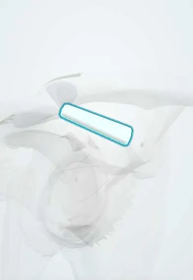
The coracoacromial ligament forms part of the roof over the shoulder joint; it keeps the ball from riding up while the cuff tendons glide beneath.
Opens in the Durgical app, no sign-up needed.
What it is
A flat band between the coracoid and the acromion that forms the roof over the rotator cuff.
What it does
Keeps the humeral head from riding up; the cuff tendons glide under it.
Key facts
- Attaches
- Lateral border of the coracoid to the tip of the acromion
- Function
- Roof of the coracoacromial arch; blocks upward displacement of the head
- Tight in
- Not joint-dependent (static roof)
Relations
- Medial
- Lateral border of the coracoid process, above the origin of the coracohumeral ligament
- Lateral
- Tip and front undersurface of the acromion
- Below
- Subacromial bursa, then the supraspinatus tendon and the humeral head
- Above
- Lateral clavicle and the AC joint above and behind it
- In front
- Front deltoid, which covers it
How to find it
Locate the coracoid deep in the outer part of the infraclavicular hollow and the front edge of the acromion: the ligament spans the gap between them under the anterior deltoid and is not felt itself. In the subacromial space at arthroscopy it is the band on the underside of the front of the acromion.
Examination
- Neer impingement sign: passive full forward elevation of the internally rotated arm with the scapula held; pain suggests impingement under the arch
- Hawkins–Kennedy test: shoulder and elbow at 90°, then forced internal rotation, driving the cuff under the coracoacromial ligament; pain suggests impingement
On imaging
The supraspinatus outlet view (a scapular Y view with slight caudal tilt) shows the arch and the shape of the acromion (flat, curved or hooked); an acromial spur pointing along the ligament is a classic finding. Ultrasound shows the bursa and cuff gliding under it.
Common problems
- Part of the tight 'roof' that can rub on the cuff tendons (subacromial impingement).
- Kept or repaired in some surgeries because it stops the humeral head riding up.
How it is diagnosed
Pain when raising the arm between about 60 and 120 degrees and impingement tests. X-rays show the acromion shape; ultrasound or MRI show the cuff and bursa.
Treatment
- Physical therapy, activity changes and sometimes a steroid injection in the bursa.
- Arthroscopic bursa removal, sometimes with shaving of the acromion, often during a rotator cuff repair.
Clinical notes
- Subacromial decompression releases or partly resects the ligament and smooths the acromion; the acromial branch of the thoracoacromial artery runs on it and bleeds when it is cut
- In older AC reconstructions it is transferred to the end of the clavicle to replace the coracoclavicular ligaments (Weaver–Dunn procedure)
Anatomical variants
- Its shape varies: Y-shaped with two bands, broad, quadrangular or a single band; the two-band form is the one most often described
Related conditions
Test yourself
Think of the answer, then tap the question to see it.
Which structures form the coracoacromial arch?
The coracoacromial ligament with the acromion and the coracoid process, the roof over the subacromial bursa and supraspinatus tendon.
Why is the coracoacromial ligament preserved in a massive irreparable rotator cuff tear?
It is the last barrier against anterosuperior escape of the humeral head.
Which artery bleeds when the coracoacromial ligament is cut in subacromial decompression?
The acromial branch of the thoracoacromial artery, which runs on the ligament.
Which tests drive the rotator cuff under the coracoacromial arch?
The Neer impingement sign and the Hawkins–Kennedy test; pain suggests subacromial impingement.
Educational information, not medical advice. For any symptom, see a health professional.
Its names
- Official Latin (TA2)
- ligamentum coracoacromiale TA2 1740
- Official Spanish (DeCS)
- Ligamento coracoacromial
- English (TA2)
- Coracoacromial ligament
- Also called
- coraco-acromial ligament
Related
Sources: Terminologia Anatomica (TA2); Anatomy, Shoulder and Upper Limb, Acromioclavicular Joint; Anatomy of the Human Body (20th ed.): Surface Anatomy of the Upper Extremity (Lea & Febiger (Gray's Anatomy, 1918), 1918) · adapted, Public domain.
Spotted a mistake?We are always improving for the community. Tell us what is wrong and our team will review it.Report a mistake
Representative model, for educational purposes only.