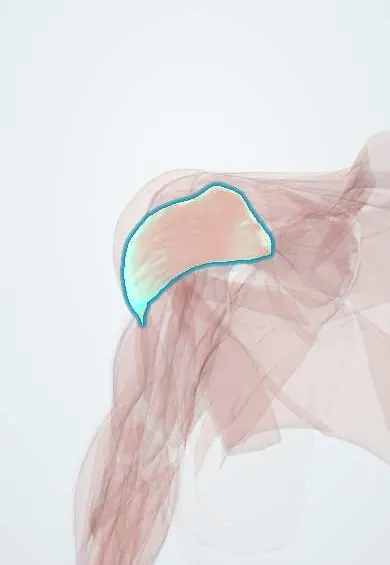
Front deltoid !
The front deltoid is the front part of the shoulder's rounded muscle; it raises the arm forward and turns it inward.
Official name: Deltoid, anterior (clavicular) part
Opens in the Durgical app, no sign-up needed.
What it is
The front part of the deltoid; it lifts the arm forward. Surgeons work beside it in the deltopectoral approach.
What it does
Raises the arm forward and turns it inward.
Key facts
- Origin
- Lateral third of the clavicle
- Insertion
- Deltoid tuberosity of the humerus
- Action
- Flexes and medially rotates the arm
- Nerve
- Axillary nerve (C5–C6)
- Blood supply
- Posterior circumflex humeral artery, thoracoacromial artery (deltoid branch)
Relations
- Medial
- Clavicular head of the pectoralis major across the deltopectoral groove, with the cephalic vein
- Deep
- Coracoid process, the conjoint tendon of coracobrachialis and the short head of biceps, the subacromial bursa and the long biceps tendon in its groove
- Lateral
- Middle deltoid
- Above
- Lateral third of the clavicle; the clavipectoral triangle at its upper medial corner
How to find it
Raising the arm forward against resistance, elbow bent, makes the front of the shoulder bulge below the outer clavicle. The groove at its medial edge, toward the chest, marks the cephalic vein.
Examination
- Resisted forward flexion at about 90°: weakness, with intact pectoral and biceps strength, points to the axillary nerve or the C5 root
On imaging
Ultrasound and MRI show it at the front of the shoulder, lateral to the deltopectoral groove and the cephalic vein. Strain shows as oedema and denervation as atrophy with fatty change; atrophy limited to the front part after a deltoid-splitting approach suggests injury to the anterior branch of the axillary nerve. Radiographs do not assess it.
Common problems
- Weakness or wasting when the axillary nerve is injured (shoulder dislocation, humerus fracture).
- In a reverse shoulder replacement it becomes the main muscle that lifts the arm.
How it is diagnosed
Exam of shoulder lifting strength and of the skin feeling over the outer shoulder; EMG if a nerve injury is suspected.
Treatment
- Physical therapy to strengthen it.
- Its strength is what makes a reverse shoulder replacement work.
Clinical notes
- In the deltopectoral approach the cephalic vein is usually kept and retracted with the deltoid; the deltoid origin is spared to protect elevation
- After an axillary nerve injury, some forward flexion persists through the clavicular head of the pectoralis major, the coracobrachialis and the biceps, which can hide the deficit
- In distal clavicle and AC joint surgery its origin is lifted with the trapezius insertion as one deltotrapezial layer and repaired afterward
Related conditions
Test yourself
Think of the answer, then tap the question to see it.
Why is the deltopectoral approach an internervous plane?
It runs between the front deltoid (axillary nerve) and the pectoralis major (pectoral nerves).
How can a deltoid-splitting incision paralyse the front deltoid?
A low split of the middle deltoid can cut the anterior branch of the axillary nerve, which runs forward deep to the muscle.
Why can forward flexion persist after an axillary nerve injury despite a paralysed front deltoid?
The clavicular head of the pectoralis major, the coracobrachialis and the biceps still flex the arm, which can hide the deficit.
Educational information, not medical advice. For any symptom, see a health professional.
Its names
- Official Latin (TA2)
- pars clavicularis musculi deltoidei TA2 2453
- Official Spanish (DeCS)
- Porción clavicular del músculo deltoides
- English (TA2)
- Deltoid, anterior (clavicular) part
- Also called
- anterior deltoid
Related
Sources: Terminologia Anatomica (TA2); Anatomy, Shoulder and Upper Limb, Deltoid Muscle.
Spotted a mistake?We are always improving for the community. Tell us what is wrong and our team will review it.Report a mistake
Representative model, for educational purposes only.