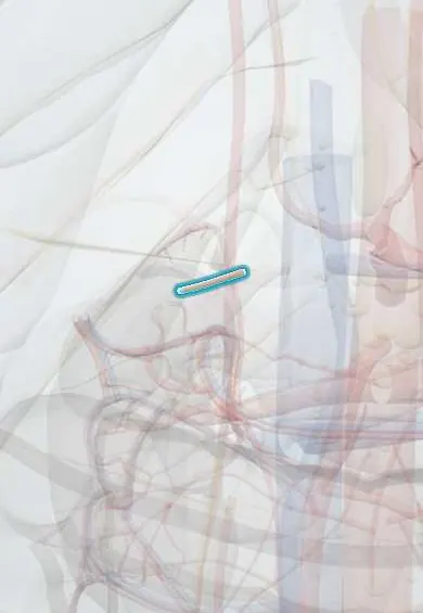
Cardiovascular system · Abdomen
Cystic artery !
The cystic artery is the small artery that feeds the gallbladder; it is found and divided in the hepatocystic (Calot) triangle when the gallbladder is removed.
Opens in the Durgical app, no sign-up needed.
What it is
A small artery, usually from the right hepatic artery, to the gallbladder; it runs through the triangle of Calot.
What it does
Supplies the gallbladder. It is clipped and cut in a cholecystectomy.
Key facts
- Origin
- Usually right hepatic artery, inside hepatocystic (Calot) triangle (varies: left or common hepatic, gastroduodenal; may be double)
- Branches
- Superficial (anterior) and deep (posterior) branches
- Supplies
- Gallbladder and cystic duct
Relations
- Course
- Usually leaves the right hepatic artery inside the hepatocystic triangle, runs above the cystic duct to the neck of the gallbladder and divides on its surface
- Below
- Cystic duct
- Medial
- Common hepatic duct, with the right hepatic artery passing behind it
- Above
- Underside of the liver and the gallbladder bed
- Superficial
- Peritoneum over the triangle and the cystic lymph node
How to find it
Found at laparoscopic cholecystectomy: with the gallbladder fundus pushed up over the liver and the neck pulled out to the side, the triangle opens and the artery is dissected free above and behind the cystic duct, often next to the cystic lymph node.
On imaging
Usually not seen on routine imaging. Color Doppler may show flow in the gallbladder wall; in acute cholecystitis, more wall flow can add to the main signs: stones, a thick wall, pericholecystic fluid and a positive sonographic Murphy sign. CT angiography maps it before surgery when variant anatomy is a concern, and after cholecystectomy it can show bleeding or a pseudoaneurysm from the stump.
Common problems
- Gallstones: very common (more in women, after pregnancy, with obesity, and in some Latin American populations); many cause no symptoms.
- Biliary colic: pain under the right ribs after meals when a stone blocks the outlet.
- Cholecystitis: an inflamed, infected gallbladder with fever.
How it is diagnosed
Ultrasound; blood tests.
Treatment
- Removal of the gallbladder by laparoscopy (cholecystectomy) for symptomatic stones.
Clinical notes
- It is clipped and divided only after the critical view of safety; a vessel thicker than expected may be the right hepatic artery
- A slipped clip or a torn deep branch can bleed enough to force conversion to open surgery
- In acute cholecystitis, raised pressure in the obstructed gallbladder reduces flow through it; gangrene and perforation can follow
- A cystic artery pseudoaneurysm is a rare cause of bleeding into the bile ducts (haemobilia) after cholecystitis or cholecystectomy
Anatomical variants
- Double cystic artery: both vessels are identified and divided
- An origin outside the triangle (gastroduodenal, left or common hepatic artery) makes it run in front of the bile duct or below the cystic duct
- A very short cystic artery from a looping right hepatic artery (caterpillar hump)
Related conditions
Test yourself
Think of the answer, then tap the question to see it.
Where does the cystic artery usually arise?
From the right hepatic artery, inside the hepatocystic (Calot) triangle, running above the cystic duct to the neck of the gallbladder.
What are the boundaries of the hepatocystic (Calot) triangle?
The cystic duct below, the common hepatic duct medially and the underside of the liver above; it contains the cystic artery and the cystic lymph node.
What is the critical view of safety in cholecystectomy?
The triangle is cleared and the lower gallbladder lifted off the liver until only two structures, the cystic duct and cystic artery, enter the gallbladder.
What may a cystic artery thicker than expected actually be?
The right hepatic artery, for example a looping right hepatic artery (caterpillar hump) that gives off a very short cystic artery.
Educational information, not medical advice. For any symptom, see a health professional.
Its names
- Official Latin (TA2)
- arteria cystica TA2 4230
- Official Spanish (DeCS)
- Arteria cística
- English (TA2)
- Cystic artery
- Also called
- arteria vesicae felleae
Related
Sources: Terminologia Anatomica (TA2); Anatomy, Abdomen and Pelvis: Celiac Trunk.
Spotted a mistake?We are always improving for the community. Tell us what is wrong and our team will review it.Report a mistake
Representative model, for educational purposes only.