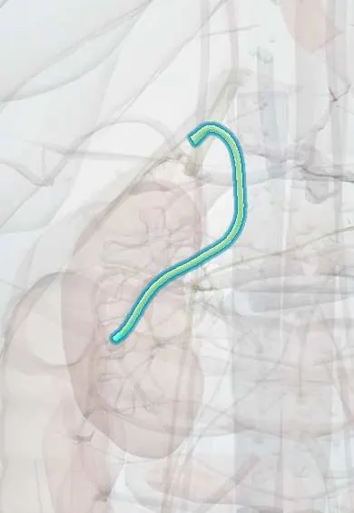
Common bile duct !
The common bile duct (choledochus) carries bile from the liver and gallbladder down to the duodenum; a stone blocking it causes jaundice.
Official name: Bile duct
Opens in the Durgical app, no sign-up needed.
What it is
The main bile duct, running behind the duodenum and through the head of the pancreas to open into the duodenum.
What it does
Carries bile from the liver and gallbladder to the intestine.
Key facts
- Where
- About 6–8 cm: supraduodenal (free edge of lesser omentum, right of hepatic artery, in front of portal vein), retroduodenal, pancreatic, intraduodenal
- Function
- Carries bile to the duodenum
- Drains
- Common hepatic duct and cystic duct
- Into
- Joins main pancreatic duct at hepatopancreatic ampulla, opening on major duodenal papilla (D2)
Relations
- Course
- Down the free edge of the lesser omentum, behind the first part of the duodenum, then in a groove on (or through) the back of the head of the pancreas, entering the second part of the duodenum obliquely
- Behind
- Portal vein in the hepatoduodenal ligament, with the epiploic foramen behind the free edge; lower down, the inferior vena cava behind the pancreatic head
- Medial
- Proper hepatic artery on its left above; the gastroduodenal artery nearby behind the first part of the duodenum
- In front
- First part of the duodenum, then pancreatic tissue where it runs through the head of the pancreas
How to find it
It cannot be felt. In open surgery it lies in the free edge of the lesser omentum, which can be held between finger and thumb through the epiploic foramen (as in the Pringle maneuver); a Kocher maneuver, lifting the duodenum and the pancreatic head, exposes its lower part.
On imaging
Ultrasound measures its width and shows dilation above a block, but often misses distal stones hidden by bowel gas. MRCP shows stones as dark filling defects within bright bile; endoscopic ultrasound picks up small stones well; ERCP is kept for treatment (stone removal, sphincterotomy, stent).
Common problems
- A gallstone stuck in it: jaundice, pain, and sometimes infection (cholangitis) or pancreatitis.
- Injury during cholecystectomy (rare but serious).
How it is diagnosed
Blood tests, ultrasound, MRI (MRCP).
Treatment
- Stones removed by ERCP (an endoscope through the mouth to the duodenum), then usually cholecystectomy.
Clinical notes
- Obstructive jaundice: dark urine, pale stools and itching; stones tend to give painful, fluctuating jaundice, tumours painless, progressive jaundice
- Ascending cholangitis is obstruction plus infection; draining the duct, usually by ERCP, is the key step together with antibiotics
- Choledochal cysts are congenital dilations of the duct (Todani classification) that raise the risk of stones, cholangitis and cancer
Anatomical variants
- Its union with the main pancreatic duct varies: a common channel (most often), separate openings on the same papilla, or rarely separate papillae
- A long common channel outside the sphincter (pancreaticobiliary maljunction) is linked to choledochal cysts and gallbladder cancer
Development
Comes from the stalk of the hepatic diverticulum; as the duodenum rotates, its opening moves with the ventral pancreatic bud from the front to the posteromedial wall of the duodenum.
Related conditions
- Gallstones (cholelithiasis)
- Choledocholithiasis
- Acute cholangitis
- Pancreatic cancer
- Gallbladder cancer
- Primary sclerosing cholangitis
- Cholangiocarcinoma (bile duct cancer)
- Choledochal cyst
- Liver fluke infections (fascioliasis, clonorchiasis, opisthorchiasis)
- Biliary atresia
Test yourself
Think of the answer, then tap the question to see it.
What is Charcot triad?
Fever with chills, jaundice and right upper quadrant pain: ascending cholangitis; Reynolds pentad adds low blood pressure and confusion.
What is the course of the common bile duct?
Down the free edge of the lesser omentum, behind D1, through or behind the head of the pancreas, joining the pancreatic duct to open on the major papilla in D2.
How does jaundice from a common bile duct stone usually differ from a tumour?
Stones tend to give painful, fluctuating jaundice; tumours painless, progressive jaundice.
Which test shows common bile duct stones without instrumenting the duct?
MRCP, showing stones as dark filling defects in bright bile; ERCP is kept for treatment.
Educational information, not medical advice. For any symptom, see a health professional.
Its names
- Official Latin (TA2)
- ductus biliaris TA2 3103
- Official Spanish (DeCS)
- Conducto colédoco DeCS D003135
- English (TA2)
- Bile duct
Related
Sources: Terminologia Anatomica (TA2); DeCS; Anatomy, Abdomen and Pelvis: Biliary Ducts.
Spotted a mistake?We are always improving for the community. Tell us what is wrong and our team will review it.Report a mistake
Representative model, for educational purposes only.