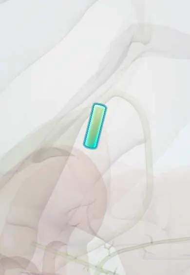
Cystic duct !
The cystic duct is the short tube that connects the gallbladder to the main bile duct; a stone stuck in it causes biliary colic and cholecystitis.
Opens in the Durgical app, no sign-up needed.
What it is
The short duct from the gallbladder neck to the bile duct.
What it does
Lets bile in and out of the gallbladder. In a cholecystectomy it is clipped and cut only after it is clearly identified (critical view of safety).
Key facts
- Where
- About 3–4 cm, from gallbladder neck to common hepatic duct, in hepatoduodenal ligament
- Attaches
- Mucosal spiral valve (of Heister); one border of hepatocystic (Calot) triangle with common hepatic duct and liver edge
- Function
- Carries bile into and out of the gallbladder
- Into
- Joins common hepatic duct to form common bile duct
Relations
- Course
- From the gallbladder neck it runs down, back and to the left in the hepatoduodenal ligament (free edge of the lesser omentum) to join the common hepatic duct, usually above the duodenum
- Above
- Cystic artery, usually above it and roughly parallel inside the hepatocystic triangle, with the cystic lymph node close by
- Medial
- Common hepatic duct, the medial side of the hepatocystic (Calot) triangle; the right hepatic artery often passes just behind it
How to find it
At laparoscopic cholecystectomy the gallbladder neck (Hartmann pouch) is pulled out to the side, and the duct is found where the neck narrows into a tube heading toward the common hepatic duct. It is clipped and divided only after the critical view of safety shows it clearly entering the gallbladder.
On imaging
MRCP shows it as a thin, often tortuous bright tube from the gallbladder neck and maps where and how it joins before surgery. On hepatobiliary scintigraphy (HIDA), tracer that reaches the bowel while the gallbladder never fills means the cystic duct is blocked, as in acute cholecystitis.
Common problems
- Gallstones: very common (more in women, after pregnancy, with obesity, and in some Latin American populations); many cause no symptoms.
- Biliary colic: pain under the right ribs after meals when a stone blocks the outlet.
- Cholecystitis: an inflamed, infected gallbladder with fever.
How it is diagnosed
Ultrasound; blood tests.
Treatment
- Removal of the gallbladder by laparoscopy (cholecystectomy) for symptomatic stones.
Clinical notes
- Dissection in the hepatocystic triangle stays close to the gallbladder to protect the common hepatic duct and right hepatic artery; a low, parallel junction makes the two ducts easy to confuse
- A cystic stump leak shows as bile collecting under the liver days after surgery and is usually managed endoscopically with a biliary stent
Anatomical variants
- Its junction with the common hepatic duct varies: low and parallel (both in one sheath), spiralling behind the common hepatic duct to join its left side, or high near the hilum
- It may drain into the right hepatic duct or an aberrant right posterior duct, which puts that duct at risk during cholecystectomy
- Very short or absent, with the gallbladder opening almost straight onto the bile duct
Development
The gallbladder and cystic duct grow from a small caudal bud of the hepatic diverticulum, an outgrowth of the foregut endoderm; a doubled bud explains the rare double gallbladder with two cystic ducts.
Related conditions
Test yourself
Think of the answer, then tap the question to see it.
Why does a stone impacted in the cystic duct not usually cause jaundice?
The bile ducts stay open; the stone causes biliary colic and then acute cholecystitis.
What are the borders of the hepatocystic (Calot) triangle?
The cystic duct, the common hepatic duct and the edge of the liver; the cystic artery usually runs inside it.
What is the most common source of bile leak after cholecystectomy?
The cystic duct stump (slipped clip or necrosis), usually managed endoscopically with a biliary stent.
Educational information, not medical advice. For any symptom, see a health professional.
Its names
- Official Latin (TA2)
- ductus cysticus TA2 3101
- Official Spanish (DeCS)
- Conducto cístico DeCS D003549
- English (TA2)
- Cystic duct
Related
Sources: Terminologia Anatomica (TA2); DeCS; Anatomy, Abdomen and Pelvis: Biliary Ducts; 23.6 Accessory Organs in Digestion: The Liver, Pancreas, and Gallbladder (OpenStax (Rice University), 2022) · adapted, CC BY 4.0.
Spotted a mistake?We are always improving for the community. Tell us what is wrong and our team will review it.Report a mistake
Representative model, for educational purposes only.