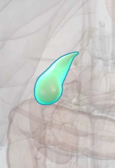
Acute cholecystitis !
Acute cholecystitis is inflammation of the gallbladder, usually because a gallstone blocks its outlet; it causes lasting pain in the upper right belly.
Updated 2026-10-29
Structures involved
Tap one to see what it is and open it in 3D.
In histology
In the clinical exam
- Abdominal regions and quadrants: what lies under each
- Abdominal palpation: light and deep, guarding and masses
- Murphy sign
- Rebound tenderness (Blumberg sign)
- Acute abdomen and peritoneal irritation
In physiology
Why it happens
- Patterns of acute inflammation: serous, fibrinous, purulent, abscess, ulcer
- Bile and the enterohepatic circulation
What it is
Acute inflammation of the gallbladder, in about nine out of ten cases caused by a gallstone blocking the cystic duct (calculous cholecystitis).
Who gets it
The most common complication of gallstones; more frequent in women and with age. Gallstone disease is common in Latin America, and cholecystectomy is one of the most frequent operations in Colombia.
Causes
- Gallstone impacted in the cystic duct or gallbladder neck (calculous)
- Acalculous cholecystitis: critical illness, major surgery, burns, prolonged fasting or parenteral nutrition
- Rarely: infections or ischaemia of the gallbladder
Risk factors
- Known gallstones and previous biliary colic
- Female sex, pregnancy and older age
- Obesity, rapid weight loss
- Diabetes
- Critical illness (acalculous form)
Pathophysiology
A stone blocks the cystic duct, so the gallbladder distends and its wall is irritated by concentrated bile. Inflammation follows, with secondary bacterial infection in many cases. Raised pressure can compromise blood flow, leading to gangrene, perforation or empyema.
Classification
- Tokyo guidelines severity: grade I (mild), grade II (moderate, with marked local inflammation), grade III (severe, with organ dysfunction)
- Calculous or acalculous
- Complicated forms: gangrenous, emphysematous, empyema, perforation
Symptoms and signs
- Constant right upper quadrant or epigastric pain lasting more than several hours, often after a fatty meal
- Pain radiating to the right shoulder or back
- Fever, nausea and vomiting
- Murphy sign: the patient stops breathing in when the examiner presses under the right costal margin
- Tender, sometimes palpable, gallbladder
Diagnosis
- Clinical
- The Tokyo guidelines combine local signs (Murphy sign, right upper quadrant mass or pain), systemic signs (fever, raised C-reactive protein or white cells) and imaging. Criteria have been updated over time.
- Tests
- Blood count, C-reactive protein, liver tests, bilirubin and lipase. Mild rises in liver tests are common; marked rises suggest a stone in the common bile duct.
- Imaging
- Ultrasound is first line: stones, thickened wall, pericholecystic fluid and a sonographic Murphy sign. Cholescintigraphy (HIDA) is the most sensitive test when ultrasound is unclear; CT helps with complications.
Differential diagnosis
- Biliary colic without inflammation
- Acute cholangitis or choledocholithiasis
- Acute pancreatitis
- Peptic ulcer disease or perforation
- Right lower lobe pneumonia
- Acute hepatitis or inferior myocardial infarction
Goal of treatment
Control inflammation and infection and remove the gallbladder early to prevent complications and recurrence.
Treatment
- Acute
- Fasting, intravenous fluids, analgesia (including non-steroidal anti-inflammatory drugs or opioids) and antiemetics.
- Medical
- Antibiotics covering gut Gram-negative and anaerobic bacteria, according to severity and local resistance.
- Surgical
- Early laparoscopic cholecystectomy, ideally during the same admission, is preferred for patients fit for surgery.
- Procedure
- Percutaneous cholecystostomy (gallbladder drainage) for very sick or high-risk patients who cannot have surgery yet.
- Follow-up
- Close watch for organ dysfunction and for stones in the common bile duct.
Complications
- Gangrene and perforation of the gallbladder
- Empyema or pericholecystic abscess
- Emphysematous cholecystitis (gas-forming infection)
- Cholecystoenteric fistula and gallstone ileus
- Sepsis
- Bile duct injury during cholecystectomy
Prognosis
Most patients recover well after early cholecystectomy. Severe (grade III) disease, acalculous cholecystitis and older age carry higher mortality.
Prevention
- Elective cholecystectomy is considered for people with recurrent symptomatic gallstones
- Healthy weight and avoiding very rapid weight loss
Test yourself
Think of the answer, then tap the question to see it.
What is Murphy sign?
Inspiratory arrest when the examiner presses below the right costal margin as the patient breathes in, because the inflamed gallbladder meets the hand.
What is the first-line imaging test in suspected acute cholecystitis?
Abdominal ultrasound: gallstones, thickened gallbladder wall, pericholecystic fluid and a sonographic Murphy sign.
What do the Tokyo guidelines provide for acute cholecystitis?
Diagnostic criteria (local signs, systemic signs, imaging) and a severity grade I–III that guides management.
What is the preferred definitive treatment in a fit patient?
Early laparoscopic cholecystectomy, ideally during the same admission.
In whom is acalculous cholecystitis typically seen?
Critically ill patients: after major surgery, trauma, burns, sepsis or long fasting with parenteral nutrition.
Related conditions
- Gallstones (cholelithiasis)
- Choledocholithiasis
- Acute cholangitis
- Acute pancreatitis
- Peptic ulcer disease
- Amoebic liver abscess
- Gallbladder cancer
Educational information, not medical advice. For any symptom, see a health professional.
Sources: Acute Cholecystitis (StatPearls (NCBI Bookshelf), 2025).
Spotted a mistake?We are always improving for the community. Tell us what is wrong and our team will review it.Report a mistake
Representative model, for educational purposes only.