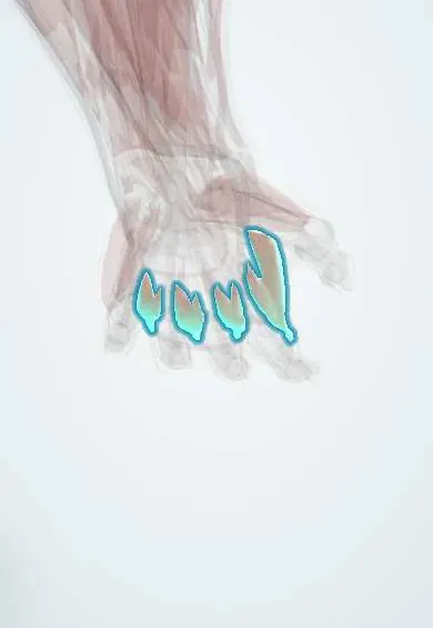
Dorsal interossei !
The dorsal interossei are four muscles between the palm bones; they spread the fingers apart from the middle finger.
Opens in the Durgical app, no sign-up needed.
What it is
Four muscles between the metacarpals, felt on the back of the hand.
What it does
Spread the fingers apart and help the lumbricals.
Key facts
- Origin
- Adjacent sides of two metacarpals (two heads each)
- Insertion
- Extensor expansions and bases of the proximal phalanges of digits 2–4
- Action
- Abduct the fingers (DAB); flex MCP, extend IP
- Nerve
- Deep branch of the ulnar nerve (C8–T1)
- Blood supply
- Dorsal metacarpal arteries, deep palmar arch
Relations
- Behind
- The extensor digitorum tendons with the juncturae, the dorsal fascia of the hand and the dorsal venous network
- In front
- The palmar interossei, then the deep palmar arch and the deep branch of the ulnar nerve; the adductor pollicis in front of the first
- Contents
- Between the two heads of the first, the radial artery on its way from the back of the hand into the palm
How to find it
The first is seen and felt as the bulge on the back of the first web space when the index is pushed toward the thumb against resistance; the others lie between the metacarpals on the back of the hand.
Examination
- Resisted index abduction (first dorsal interosseous): weakness or a hollow first web points to the ulnar nerve, or to the C8–T1 roots or lower trunk
- Egawa sign: with the hand flat, the middle finger cannot be rocked from side to side, indicating weakness of the 2nd and 3rd dorsal interossei (ulnar palsy)
- Bunnell–Littler test: the PIP joint bends less when the MCP joint is held straight than when it is bent, indicating intrinsic tightness
On imaging
On MRI and ultrasound they fill the spaces between the metacarpals, seen best from the back of the hand. They are not imaged on their own for injury. The classic finding is wasting with ulnar nerve palsy, most visible in the first web space; MRI shows it as oedema early and fatty atrophy later. Electrodiagnostic studies, not imaging, confirm the nerve lesion.
Clinical notes
- Ulnar claw hand reflects loss of the interossei and ulnar lumbricals: MCP joints hyperextend and IP joints flex
- Intrinsic tightness (after crush injury, swelling or rheumatoid arthritis) pulls the fingers toward the intrinsic-plus position and can lead to swan-neck deformity
- The interosseous compartments of the hand are released through dorsal incisions over the 2nd and 4th metacarpals in hand compartment syndrome
Anatomical variants
- Classically all bipennate, but about a quarter were unipennate in one dissection study
- With a Martin–Gruber anastomosis, median fibres reach the first dorsal interosseous through the ulnar nerve, so it may be spared in a high ulnar lesion
Development
The fingers separate in weeks 6–7 as cells between the digital rays die; the interossei form in the tissue left between the metacarpals. Their C8–T1 supply means a lower plexus birth injury (Klumpke palsy) wastes them and claws the hand.
Memory aid
DAB: Dorsal interossei ABduct, measured from the middle finger, which has two of them because it moves both ways.
Related conditions
Test yourself
Think of the answer, then tap the question to see it.
What do the dorsal interossei do, and around which axis?
They abduct the fingers from the axis of the middle finger; the middle finger has two, the thumb and little finger none.
What is the most visible early sign of an ulnar nerve lesion in the hand?
Wasting of the first dorsal interosseous in the first web space.
What is the Egawa sign?
With the hand flat, the middle finger cannot be rocked from side to side: weak 2nd and 3rd dorsal interossei (ulnar palsy).
Educational information, not medical advice. For any symptom, see a health professional.
Its names
- Official Latin (TA2)
- musculi interossei dorsales manus TA2 2533
- Official Spanish (DeCS)
- Músculos interóseos dorsales de la mano
- English (TA2)
- Dorsal interossei
Related
- Ulnar nerve in the hand
- Deep palmar arch
- 1st metacarpal
- 2nd metacarpal
- 3rd metacarpal
- 4th metacarpal
- 5th metacarpal
- Proximal phalanx, middle finger
Sources: Terminologia Anatomica (TA2); Anatomy, Shoulder and Upper Limb, Hand Dorsal Interossei Muscle; 8.5 Development of the Appendicular Skeleton (OpenStax (Rice University), 2022) · adapted, CC BY 4.0.
Spotted a mistake?We are always improving for the community. Tell us what is wrong and our team will review it.Report a mistake
Representative model, for educational purposes only.