Joints and ligaments · Lower limb
Iliofemoral ligament !
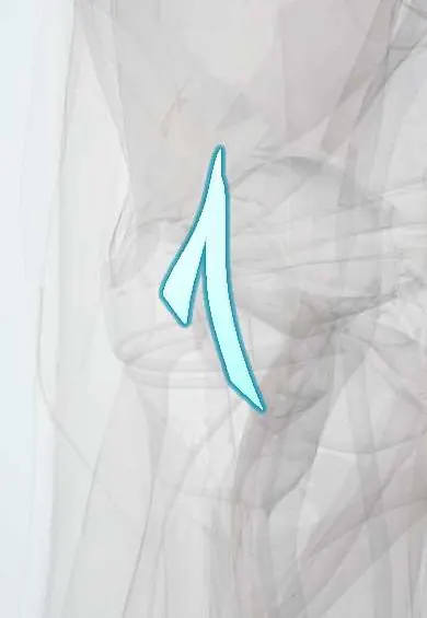
The iliofemoral ligament is a strong Y-shaped band at the front of the hip; it stops the hip bending too far backward.
Opens in the Durgical app, no sign-up needed.
What it is
A thick, Y-shaped ligament on the front of the hip capsule, from the front of the pelvis to the front of the femur. It is one of the strongest ligaments in the body.
What it does
Stops the hip from bending too far backward, so you can stand upright with little muscle effort. It also limits the thigh turning outward.
Key facts
- Attaches
- Anterior inferior iliac spine and acetabular rim to intertrochanteric line (inverted Y, of Bigelow)
- Function
- Strongest ligament of body; prevents hip hyperextension, letting us stand with little muscle effort
- Tight in
- Hip extension (and external rotation)
Relations
- In front
- Iliopsoas tendon with the iliopsoas bursa between, which may communicate with the joint; the femoral nerve and vessels lie further in front
- Above
- Reflected head of rectus femoris along the acetabular rim, and gluteus minimus laterally
- Medial
- Pubofemoral ligament, with a thin capsular area between them under the iliopsoas tendon
- Deep
- Femoral head and neck, with the circular fibres of the zona orbicularis
How to find it
Not palpable: the hip joint lies deep under the iliopsoas and the rectus femoris. In an anterior approach it is the thick band on the front of the capsule, seen once those muscles are retracted, running from just below the anterior inferior iliac spine to the intertrochanteric line; arthroscopic capsulotomy cuts across it between the portals.
Examination
- Anterior apprehension test: hip extended and externally rotated; apprehension or anterior pain suggests anterior capsular laxity (microinstability)
- Hip dial test: lying supine, the straight leg is rotated inward and released; if it rolls out well past neutral without a firm end point, the anterior capsule may be lax
On imaging
Not seen on X-ray. MRI or MR arthrography shows it as the thick dark anterior part of the capsule on axial oblique images; thinning or a defect may be seen after hip arthroscopy.
Common problems
- Stretched or torn in a hip dislocation, which usually takes a large force.
- Partial tears or looseness can cause a feeling of hip instability, sometimes after hip arthroscopy.
- In arthritis it can become tight and limit hip movement.
How it is diagnosed
Physical exam; X-rays; MRI or MR arthrogram to see the capsule and ligaments.
Treatment
- Physical therapy to strengthen the muscles around the hip.
- Surgical repair or tightening of the capsule and ligament when instability persists.
Clinical notes
- Arthroscopic capsulotomy cuts across it to reach the joint; repairing the capsule afterwards helps prevent instability
- In posterior dislocation the leg looks short, flexed, adducted and internally rotated, with the intact ligament tethering the femur in front
Related conditions
Test yourself
Think of the answer, then tap the question to see it.
Where does the iliofemoral ligament attach, and what shape does it have?
From the anterior inferior iliac spine and acetabular rim to the intertrochanteric line, as an inverted Y (of Bigelow).
Which movement does the iliofemoral ligament prevent, and what does that allow?
Hip hyperextension; it lets us stand upright with little muscle effort.
What role does the iliofemoral ligament play in posterior hip dislocation?
It usually stays intact and acts as a hinge, which reduction manoeuvres such as Allis and Bigelow use.
How do people with paraplegia stand using the iliofemoral ligaments?
By leaning back with the hips extended, hanging on these ligaments.
Educational information, not medical advice. For any symptom, see a health professional.
Its names
- Official Latin (TA2)
- ligamentum iliofemorale TA2 1875
- Official Spanish (DeCS)
- Ligamento iliofemoral
- English (TA2)
- Iliofemoral ligament
- Also called
- y ligament of Bigelow · ligament of Bertin
Related
Sources: Terminologia Anatomica (TA2); Anatomy, Bony Pelvis and Lower Limb, Hip Joint; Anatomy of the Human Body (20th ed.): Surface Anatomy of the Lower Extremity (Lea & Febiger (Gray's Anatomy, 1918), 1918) · adapted, Public domain.
Spotted a mistake?We are always improving for the community. Tell us what is wrong and our team will review it.Report a mistake
Representative model, for educational purposes only.