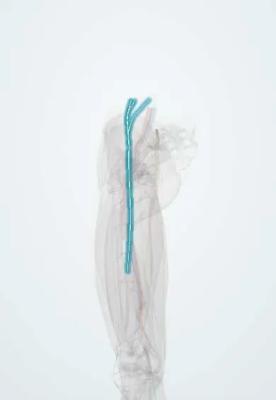
Femoral nerve !
The femoral nerve is the main nerve of the front of the thigh; it powers the quadriceps that straighten the knee and carries feeling.
Opens in the Durgical app, no sign-up needed.
What it is
A large nerve from the lower spine (lumbar plexus). It runs down beside the psoas, passes under the inguinal ligament next to the femoral artery, and branches out in the front of the thigh.
What it does
Controls the quadriceps (straightening the knee) and helps lift the thigh. It carries feeling from the front of the thigh and, through the saphenous nerve, from the inner leg.
Key facts
- Roots
- L2–L4 (posterior divisions, lumbar plexus)
- Moves
- Iliacus, pectineus, sartorius, quadriceps femoris; knee extension (knee jerk L3–L4)
- Feeling from
- Anterior and medial thigh (anterior cutaneous branches); medial leg and foot (saphenous nerve)
Relations
- Course
- Forms within the psoas major, emerges from its lateral border, descends in the groove between the psoas and iliacus beneath the fascia iliaca, passes under the inguinal ligament lateral to the femoral sheath and splits in the femoral triangle into anterior and posterior divisions
- Medial
- The femoral artery, then the femoral vein and the femoral canal, all inside the femoral sheath; above the inguinal ligament the psoas major separates it from the external iliac artery, and under the ligament the iliopectineal arch does, so it passes through the muscular lacuna
- Deep
- The iliopsoas in the iliac fossa and in front of the hip
- Superficial
- The fascia iliaca, the inguinal ligament and, below it, the fascia lata
- Lateral
- The iliacus and, near the anterior superior iliac spine, the lateral femoral cutaneous nerve
How to find it
Below the inguinal ligament the femoral pulse is felt at the mid-inguinal point (halfway from the anterior superior iliac spine to the pubic symphysis). The nerve lies just lateral to the pulse, deep to the fascia iliaca, and is found there with ultrasound for a femoral block.
Examination
- Patellar reflex (L3–L4, mainly L4): reduced or absent in a femoral nerve or L4 root lesion
- Resisted knee extension and a single-leg step-up: quadriceps weakness; if hip adduction (obturator nerve) is also weak, the lesion is more likely at the L3–L4 roots or the lumbar plexus
- Femoral nerve stretch test (prone knee bend): anterior thigh pain when the knee is bent with the hip extended suggests irritation of an L2–L4 root
On imaging
Ultrasound below the inguinal ligament shows it as a flat, honeycomb-like structure lateral to the femoral artery, under the fascia iliaca; this view guides the femoral block. Above the ligament CT is first if an iliopsoas haematoma is suspected, a classic cause of femoral palsy. MRI shows denervation oedema or atrophy in the quadriceps. Radiographs do not show it.
Common problems
- Injury or stretching during hip, pelvic or abdominal surgery, or pressure from bleeding in the pelvis.
- Weak quadriceps: the knee may buckle and climbing stairs is hard.
- Numbness at the front of the thigh and the inner leg.
How it is diagnosed
Physical exam (knee reflex, thigh strength, feeling); nerve conduction studies and EMG; imaging to look for a cause such as a bleed.
Treatment
- Treating the cause, such as relieving pressure on the nerve.
- Physical therapy and a knee brace while the nerve recovers.
- Numbing medicine placed near this nerve (a nerve block) is often used to control pain after knee or hip surgery.
Clinical notes
- Iatrogenic injury from self-retaining retractor blades in abdominal and pelvic surgery, the lithotomy position, anterior hip approaches and femoral artery puncture
- Femoral and fascia iliaca blocks are used for hip fractures and thigh or knee surgery; the quadriceps weakness they cause raises the risk of falls
- Diabetic lumbosacral radiculoplexus neuropathy (diabetic amyotrophy) starts with severe thigh pain followed by quadriceps wasting and can mimic a femoral neuropathy
Development
Formed from the posterior divisions of the ventral rami of L2–L4, it supplies muscles of the dorsal muscle mass; the medial rotation of the lower limb in the seventh week carries that mass, and the nerve, to the front of the thigh.
Memory aid
NAVEL, from lateral to medial in the groin: Nerve, Artery, Vein, Empty space (femoral canal), Lymphatics.
Related conditions
Test yourself
Think of the answer, then tap the question to see it.
What are the roots of the femoral nerve?
L2–L4, posterior divisions of the lumbar plexus.
How does the femoral nerve relate to the femoral sheath under the inguinal ligament?
It is the most lateral of the femoral group and lies outside the sheath, lateral to the femoral artery.
How does hip flexion help localise a femoral nerve lesion?
A lesion above the inguinal ligament also weakens hip flexion (iliacus); below it, hip flexion is spared.
Which reflex is reduced in a femoral nerve lesion?
The patellar (knee) reflex, L3–L4, mainly L4.
What sensory territory does the femoral nerve supply?
The anterior and medial thigh, and through the saphenous nerve the medial leg and foot.
Educational information, not medical advice. For any symptom, see a health professional.
Its names
- Official Latin (TA2)
- nervus femoralis TA2 6522
- Official Spanish (DeCS)
- Nervio femoral DeCS D005267
- English (TA2)
- Femoral nerve
Related
- Inguinal ligament
- Psoas major
- Iliopsoas fascia
- Femoral artery
- Anterior cutaneous nerves of the thigh
- Saphenous nerve
- Iliacus
- Pectineus
Sources: Terminologia Anatomica (TA2); DeCS; Anatomy, Bony Pelvis and Lower Limb: Thigh Femoral Nerve; 8.5 Development of the Appendicular Skeleton (OpenStax (Rice University), 2022) · adapted, CC BY 4.0; Anatomía humana (4.ª ed.), tomo 1, cap. 70: Nervios del plexo lumbosacro.
Spotted a mistake?We are always improving for the community. Tell us what is wrong and our team will review it.Report a mistake
Representative model, for educational purposes only.