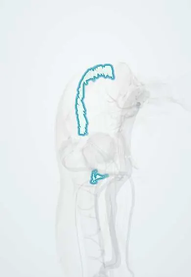
Iliopsoas tendon !
The iliopsoas tendon is the shared tendon of the psoas and iliacus; it attaches to a bump on the inner top of the femur.
Official name: Tendon of iliopsoas
Opens in the Durgical app, no sign-up needed.
What it is
The iliopsoas tendon: the shared tendon of the psoas and iliacus. It crosses the front of the hip and attaches to the lesser trochanter, a bump on the inner top of the femur.
What it does
Carries the pull of the hip flexors to the femur to lift the thigh.
Key facts
- Attaches
- Shared tendon of the psoas major and iliacus, under the inguinal ligament (muscular lacuna) and in front of the hip capsule, to the lesser trochanter; the iliopectineal bursa lies between it and the capsule
- Function
- Carries the pull of the strongest hip flexor; internal snapping hip when it flicks over the iliopectineal eminence; avulsion of the lesser trochanter in adolescents
- Tight in
- Hip extension (the Thomas test position)
Relations
- In front
- Femoral nerve under the inguinal ligament; the tendon forms the lateral part of the floor of the femoral triangle
- Behind
- Hip capsule and acetabular labrum at the front of the joint, with the iliopsoas bursa between them
- Medial
- Pectineus and the femoral artery and vein in the femoral sheath
- Below
- Lesser trochanter; the medial circumflex femoral artery passes backward between it and the pectineus
How to find it
Deep: just lateral to the femoral pulse below the inguinal ligament, the iliopsoas tightens when the seated person lifts the knee against resistance; the tendon itself is shown with ultrasound.
Examination
- Thomas test: the other knee is held to the chest to flatten the lower back; the tested thigh rising off the table shows short hip flexors
- Snapping test: moving the hip from flexion, abduction and external rotation into extension reproduces the clunk of internal snapping
- Resisted straight-leg raise (Stinchfield test): groin pain suggests a hip joint or iliopsoas source
On imaging
Dynamic ultrasound shows the tendon jumping over the pelvic brim; MRI shows tendinopathy and a distended iliopsoas bursa, which may connect with the hip joint; radiographs show lesser trochanter avulsions.
Common problems
- Strain or tendinitis from kicking, sprinting or repeated hip bending.
- Snapping hip (inside type): the tendon catches over the front of the pelvis with a clunk.
- Tight hip flexors from long sitting can add to back or hip pain.
- Uncommonly, tendon irritation after hip replacement if it rubs on the implant.
How it is diagnosed
Physical exam (pain when lifting the leg against resistance); ultrasound, which can also show the snapping during movement; MRI.
Treatment
- Rest from the painful activity, stretching and progressive strengthening with a physical therapist.
- Ultrasound-guided injection into the iliopsoas bursa or around the tendon.
- Rarely, surgery to lengthen or release the tendon when snapping or pain does not improve.
Clinical notes
- A distended iliopsoas bursa can present as a groin mass and press on the femoral vein or femoral nerve
- For persistent painful snapping the tendon can be lengthened or released at the lesser trochanter or at the level of the hip joint
- After hip replacement, an acetabular cup that stands proud of the front rim can rub on the tendon (iliopsoas impingement), giving groin pain on active hip flexion
Anatomical variants
- A bifid (double) tendon is not rare, about a quarter of hips in one series, and may contribute to snapping
Development
The lesser trochanter, where the tendon inserts, has its own secondary ossification centre that appears at about 9–10 years. Until it joins the shaft in adolescence (texts differ on the age), a forceful iliopsoas contraction can pull the apophysis off: the classic avulsion in young sprinters and kickers.
Test yourself
Think of the answer, then tap the question to see it.
Where does the iliopsoas tendon insert?
On the lesser trochanter of the femur, after passing under the inguinal ligament in front of the hip capsule.
What does an isolated lesser trochanter avulsion in an adult without major trauma suggest?
A pathological fracture through a tumour or metastasis.
What is internal snapping hip?
A clunk in the groin as the iliopsoas tendon flicks over the pelvic brim or femoral head when the bent hip is extended.
What does a positive Thomas test show?
Short hip flexors: with the other knee held to the chest, the tested thigh rises off the table.
Educational information, not medical advice. For any symptom, see a health professional.
Its names
- Official Latin (TA2)
- iliopsoas TA2 2593
- Official Spanish (DeCS)
- Tendón del psoas ilíaco DeCS D013710
- English (TA2)
- Tendon of iliopsoas
- Also called
- musculus iliopsoas · Hyrtl's muscle
Related
Sources: Terminologia Anatomica (TA2); DeCS; Anatomy, Bony Pelvis and Lower Limb: Iliopsoas Muscle; 8.5 Development of the Appendicular Skeleton (OpenStax (Rice University), 2022) · adapted, CC BY 4.0.
Spotted a mistake?We are always improving for the community. Tell us what is wrong and our team will review it.Report a mistake
Representative model, for educational purposes only.