Developmental dysplasia of the hip !
Developmental dysplasia of the hip is a baby's hip with a shallow socket, so the ball is unstable or slips partly or fully out of place.
Updated 2026-10-29
Structures involved
Tap one to see what it is and open it in 3D.
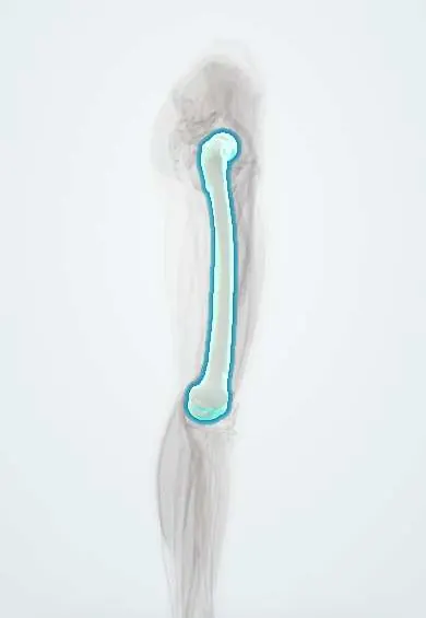
- Femur
- Hip bone
- Acetabular labrum
- Hip capsule
- Ligament of the femoral head
- Femoral head cartilage
- Acetabular cartilage
- Iliofemoral ligament
- Adductor longus
In embryology
In the clinical exam
Why it happens
What it is
Abnormal development of the infant hip in which the acetabulum is shallow and the femoral head is unstable, partly out (subluxated) or fully out (dislocated) of the socket.
Who gets it
One of the most common musculoskeletal conditions of newborns. Screening of every newborn hip is standard in paediatric care, including in Colombia; practices such as tight swaddling with straight legs increase the risk.
Causes
- Ligamentous laxity, partly from maternal hormones
- Restricted space in the uterus
- Postnatal positioning with hips straight and together
Risk factors
- Breech presentation
- Family history
- Female sex
- First-born child
- Oligohydramnios
- Tight swaddling with legs extended
Pathophysiology
Normal hip growth needs the femoral head to sit deep in the acetabulum. If the head is loose or out of place, the socket stays shallow, the labrum and capsule deform and soft tissue blocks reduction. The earlier the hip is centred, the better the socket remodels.
Classification
- Dysplastic (shallow socket), subluxatable, dislocatable or dislocated hip
- Graf classification on ultrasound
- Teratologic (fixed, prenatal) versus typical form
Symptoms and signs
- Often no symptoms in the newborn; found on screening
- Hip clunk on Ortolani or Barlow test
- Limited abduction of the hip after a few months
- Asymmetric thigh or gluteal folds and leg-length difference (Galeazzi sign)
- Painless limp or waddling gait once walking
Diagnosis
- Clinical
- Barlow test (pushes an unstable hip out) and Ortolani test (reduces a dislocated hip back in), useful in the first weeks; later, limited abduction and Galeazzi sign.
- Tests
- No laboratory tests are needed.
- Imaging
- Ultrasound before about 4–6 months, while the femoral head is still cartilage; pelvic X-ray after that age, once the ossific nucleus appears.
Differential diagnosis
- Benign hip clicks from soft tissue
- Neuromuscular conditions (cerebral palsy, spina bifida)
- Congenital short femur
- Septic arthritis of the hip
- Legg–Calvé–Perthes disease in an older child
Goal of treatment
Achieve and keep a concentric, stable reduction of the femoral head so the acetabulum develops normally, while avoiding avascular necrosis.
Treatment
- Conservative
- Pavlik harness, which holds the hips flexed and abducted, in infants up to about 6 months; criteria vary between centres.
- Procedure
- Closed reduction under anaesthesia and a spica cast when the harness fails or the child is older.
- Surgical
- Open reduction, with pelvic or femoral osteotomies in older children or residual dysplasia.
- Follow-up
- Serial ultrasound or X-ray follow-up until the hip has matured.
Complications
- Avascular necrosis of the femoral head
- Residual dysplasia
- Redislocation
- Femoral nerve palsy with the harness
- Early hip osteoarthritis in adulthood
Prognosis
Excellent when diagnosed and treated in the first months. Late diagnosis needs more invasive treatment and leads to more residual dysplasia and early arthritis.
Prevention
- Hip examination at birth and at well-child visits
- Hip-healthy swaddling that lets the legs bend and spread
- Targeted ultrasound in infants with risk factors
Test yourself
Think of the answer, then tap the question to see it.
What does the Barlow test detect?
A dislocatable hip: adduction with posterior pressure pushes the femoral head out of the acetabulum.
What does a positive Ortolani test show?
A dislocated hip that reduces with a clunk on abduction and lifting of the greater trochanter.
Which imaging is used for DDH before about 4–6 months of age, and why?
Ultrasound, because the femoral head is still cartilage; after the ossific nucleus appears, X-ray is used.
What is the first-line treatment of DDH in young infants?
A Pavlik harness, keeping the hips flexed and abducted.
What is the most feared complication of DDH treatment?
Avascular necrosis of the femoral head.
Related conditions
- Hip Osteoarthritis
- Legg-Calvé-Perthes disease
- Slipped capital femoral epiphysis
- Clubfoot (talipes equinovarus)
- Spina bifida (myelomeningocele)
- Torticollis
Educational information, not medical advice. For any symptom, see a health professional.
Sources: Developmental Dysplasia of the Hip (StatPearls (NCBI Bookshelf), 2024).
Spotted a mistake?We are always improving for the community. Tell us what is wrong and our team will review it.Report a mistake
Representative model, for educational purposes only.