Joints and ligaments · Lower limb
Hip capsule !
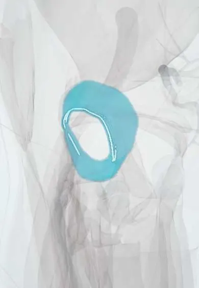
The hip capsule is a strong sleeve around the hip joint that holds the joint fluid and encloses most of the femoral neck.
Official name: Articular capsule of hip joint
Opens in the Durgical app, no sign-up needed.
What it is
A strong, thick sleeve of tissue around the hip joint, reinforced by three ligaments and lined inside by synovium.
What it does
Encloses the ball and socket, holds the joint fluid that lubricates them, and is one of the main things keeping the hip stable.
Key facts
- Where
- Encloses hip joint and most of femoral neck
- Attaches
- Acetabular rim and transverse ligament to intertrochanteric line in front; behind, junction of lateral and middle thirds of neck (about 1 cm above intertrochanteric crest)
- Function
- Strong fibrous sleeve; zona orbicularis grips the neck; reinforced by iliofemoral, pubofemoral and ischiofemoral ligaments
Relations
- In front
- Iliopsoas tendon, with the iliopsoas bursa between them; further forward the femoral nerve, femoral artery and femoral vein
- Behind
- Piriformis, the gemelli, obturator internus and quadratus femoris; the sciatic nerve lies behind them
- Above
- Reflected head of rectus femoris and gluteus minimus, which sends fibres into the capsule
- Below
- Pubofemoral ligament and the obturator externus tendon winding under and behind the neck
- Deep
- Synovial membrane, the acetabular labrum and transverse acetabular ligament at the rim, and the zona orbicularis around the neck
How to find it
Not felt directly: the joint lies deep to a point just below the middle of the inguinal ligament, lateral to the femoral pulse. Texts describe needle access to the joint from the front, lateral to the femoral vessels, or from the side above the greater trochanter, nowadays usually under ultrasound guidance.
Examination
- Log roll: rolling the relaxed, straight leg turns only the femoral head in its socket; pain or guarding points to an intra-articular source such as synovitis or infection
- Thomas test: with the other hip fully flexed, the tested thigh rising off the couch shows a fixed flexion deformity, often from a contracted anterior capsule
- Barlow and Ortolani tests in infants: a hip that slips out (Barlow) or clunks back in (Ortolani) shows the capsular laxity of developmental dysplasia
- Nélaton line: hip flexed about 45°, a line from the ASIS to the ischial tuberosity normally touches the tip of the greater trochanter; a trochanter above it suggests a femoral neck fracture or posterior dislocation
On imaging
Ultrasound along the femoral neck from the front shows an effusion as fluid lifting the capsule off the neck (anterior recess), compared with the other side, especially in children. Radiographs show effusion poorly; MRI shows synovitis, capsular thickening, tears and laxity.
Common problems
- Stiffness and pain from arthritis or inflammation of the lining (synovitis).
- Stretching or tearing in a hip dislocation, which usually needs a large force such as a car accident.
- Infection inside the joint (septic arthritis), which is an emergency.
How it is diagnosed
Physical exam; X-rays; ultrasound or MRI to see fluid or capsule damage; drawing fluid from the joint with a needle if infection is suspected.
Treatment
- Physical therapy and anti-inflammatory medicine for stiffness or synovitis.
- Dislocation: urgent reduction (putting the ball back in the socket), then protected rehabilitation.
- Surgery when needed: capsule repair during hip arthroscopy, or washing out an infected joint.
Clinical notes
- Septic arthritis: pus under pressure in the tight capsule can compress the retinacular vessels and destroy the femoral head, especially in children
- Posterior dislocation can stretch the sciatic nerve, mainly its common fibular division, and tear the retinacular vessels
- Repairing the capsule and short external rotators after hip replacement through the posterior approach lowers the dislocation rate
- Hip arthroscopy opens the capsule between portals (capsulotomy); left unrepaired, it can contribute to instability
- Nerve supply: femoral and obturator nerves in front, nerve to quadratus femoris behind (some texts add the superior gluteal); the femoral and obturator nerves also supply the knee, so hip disease can cause knee pain
Development
The newborn capsule is lax, partly from maternal hormones; this laxity is one factor in developmental dysplasia of the hip.
Related conditions
- Developmental dysplasia of the hip
- Slipped capital femoral epiphysis
- Hip dislocation
- Transient synovitis of the hip
- Polymyalgia rheumatica
Test yourself
Think of the answer, then tap the question to see it.
Why are femoral neck fractures more threatening to the femoral head than intertrochanteric fractures?
Neck fractures are intracapsular and can tear the retinacular vessels under the hip capsule; intertrochanteric fractures are extracapsular.
Where is the hip capsule weakest, and what does that explain?
Below and behind; it helps explain why most hip dislocations are posterior.
Why is septic arthritis of the hip an emergency, especially in children?
Pus under pressure in the tight hip capsule can compress the retinacular vessels and destroy the femoral head.
What does a positive Thomas test show about the hip?
A fixed flexion deformity: the tested thigh rises off the couch when the other hip is fully flexed, often from a contracted anterior capsule.
Which infant tests detect the capsular laxity of developmental dysplasia of the hip?
Barlow (the hip slips out) and Ortolani (it clunks back in).
Educational information, not medical advice. For any symptom, see a health professional.
Its names
- Official Latin (TA2)
- articulatio coxae; zona orbicularis TA2 1873
- Official Spanish (DeCS)
- Cápsula articular de la cadera DeCS D017746
- English (TA2)
- Articular capsule of hip joint
- Also called
- articulus coxae
Related
- Acetabular labrum
- Iliofemoral ligament
- Pubofemoral ligament
- Ischiofemoral ligament
- Iliopsoas bursa
- Circumflex femoral arteries
- Gluteal arteries
- Femur
Sources: Terminologia Anatomica (TA2); DeCS; Anatomy, Bony Pelvis and Lower Limb, Hip Joint; Anatomía humana (4.ª ed.), tomo 1, cap. 64: Cintura pelviana.
Spotted a mistake?We are always improving for the community. Tell us what is wrong and our team will review it.Report a mistake
Representative model, for educational purposes only.