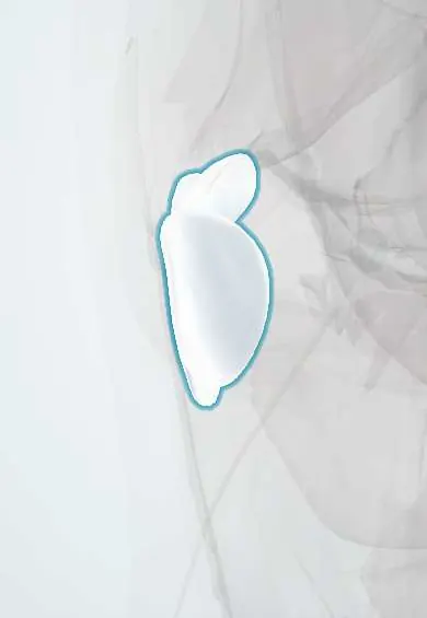
Trochanteric bursae !
The trochanteric bursae are fluid sacs over the bony point of the outer hip; they let the buttock muscles and the IT band glide.
Official name: Trochanteric bursa of gluteus maximus muscle
Opens in the Durgical app, no sign-up needed.
What it is
Fluid-filled sacs over the greater trochanter, the bony point you feel on the outer hip. They sit under the skin, the IT band, the gluteus maximus and the gluteal tendons.
What it does
Let the muscles, tendons and IT band glide smoothly over the bone.
Key facts
- Where
- Between gluteus maximus/IT band and greater trochanter; smaller bursae beneath gluteus medius and minimus
- Function
- Let gluteus maximus and IT band glide over greater trochanter; site of lateral hip pain (trochanteric bursitis)
Relations
- Superficial
- Gluteus maximus and the upper iliotibial tract; the subcutaneous trochanteric bursa lies just under the skin
- Deep
- Facets of the greater trochanter and the gluteus medius and gluteus minimus tendons
- Below
- Origin of vastus lateralis below the trochanter
How to find it
Lying on the other side with the hips and knees bent, the greater trochanter is felt as the bony prominence on the side of the hip. Tenderness over its posterolateral edge is the classic finding in greater trochanteric pain syndrome.
Examination
- Single-leg stance held for about 30 seconds that reproduces lateral hip pain points to gluteal tendinopathy
- Pain with resisted hip abduction, or with resisted external derotation from flexion and external rotation, supports a gluteal tendon source
On imaging
Ultrasound over the lateral hip shows the trochanteric facets, the gluteal tendons and any bursal fluid or thickening; MRI shows tendon tears and bursal fluid. Radiographs may show calcification or irregularity over the trochanter.
Common problems
- Trochanteric bursitis: outer hip pain, worse lying on that side, climbing stairs or after long walks. It is now usually grouped with gluteal tendinopathy as greater trochanteric pain syndrome.
- Swelling after a fall onto the hip.
- Rarely, infection.
How it is diagnosed
Physical exam (tenderness over the outer hip); ultrasound or MRI to check the tendons and the bursa.
Treatment
- Activity changes, physical therapy for the hip muscles, and anti-inflammatory medicine.
- An injection into the bursa for pain that does not settle.
- Rarely, surgery to remove the bursa (bursectomy) for long-lasting cases.
Clinical notes
- The lateral approach to the hip and the fixation of trochanteric fractures pass through the bursa
- Pain over the trochanter can also be referred from the lumbar spine or the hip joint, which examination and imaging help separate
Related conditions
Test yourself
Think of the answer, then tap the question to see it.
What does imaging of lateral hip pain more often show than a fluid-filled trochanteric bursa?
Gluteus medius and minimus tendinopathy; both are grouped as greater trochanteric pain syndrome.
Which bedside test supports gluteal tendinopathy in lateral hip pain?
Single-leg stance held for about 30 seconds that reproduces lateral hip pain.
What causes external snapping hip over the trochanteric bursae?
The iliotibial tract or the front edge of the gluteus maximus catching over the greater trochanter, often with bursal irritation.
Educational information, not medical advice. For any symptom, see a health professional.
Its names
- Official Latin (TA2)
- bursa trochanterica musculi glutei maximi; bursa trochanterica musculi glutei minimi; bursa subcutanea trochanterica TA2 2724
- Official Spanish (DeCS)
- Bolsas trocantéreas DeCS D002061
- English (TA2)
- Trochanteric bursa of gluteus maximus muscle
- Also called
- bursa trochanterica subfascialis
Related
Sources: Terminologia Anatomica (TA2); DeCS; Anatomy, Skin Bursa; Anatomy, Bony Pelvis and Lower Limb, Hip.
Spotted a mistake?We are always improving for the community. Tell us what is wrong and our team will review it.Report a mistake
Representative model, for educational purposes only.